Review
. 2008 Oct;4(5):629-36.
doi: 10.2217/14796694.4.5.629.

Emerging insights into the molecular pathogenesis of uveal melanoma

Affiliations
Review

Emerging insights into the molecular pathogenesis of uveal melanoma

Solange Landreville et al. Future Oncol. 2008 Oct.

Abstract

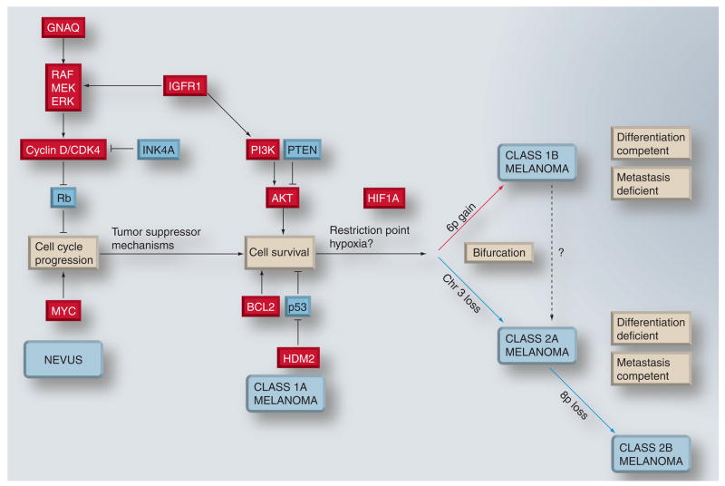
Uveal melanoma is the most common primary cancer of the eye, and often results not only in vision loss, but also in metastatic death in up to half of patients. For many years, the details of the molecular pathogenesis of uveal melanoma remained elusive. In the past decade, however, many of these details have emerged to reveal a fascinating and complex story of how the primary tumor evolves and progresses. Early events that disrupt cell cycle and apoptotic control lead to malignant transformation and proliferation of uveal melanocytes. Later, the growing tumor encounters a critical bifurcation point, where it progresses along one of two genetic pathways with very distinct genetic signatures (monosomy 3 vs 6p gain) and metastatic propensity. Late genetic events are characterized by increasing aneuploidy, most of which is nonspecific. However, specific chromosomal alterations, such as loss of chromosome 8p, can hasten the onset of metastasis in susceptible tumors. Taken together, this pathogenetic scheme can be used to construct a molecularly based and prognostically relevant classification of uveal melanomas that can be used clinically for personalized patient management.


Figures

Figure 1
Figure 1. Clinical appearance of uveal melanomas
(A) Iris melanoma. (B) Choroidal melanoma. Photograph taken with a fundus camera through the dilated pupil, showing the posterior interior of the eye.
Figure 2
Figure 2. Molecular classification of uveal melanomas based on transcriptomic and chromosomal features
(A) Unsupervised principal component analysis, showing natural clustering of uveal melanomas into four groups according to gene-expression profile and status of chromosomes 3, 6p and 8p. Class 1A – minimal aneuploidy (blue spheres); class 1B – 6p gain (green spheres); class 2A – monosomy 3 (red spheres) and class 2B – monosomy 3 and 8p loss (gray spheres). (B) Kaplan–Meier survival analysis showing that molecular classification accurately predicts metastatic death. PCA: Principle component analysis.
Figure 3
Figure 3. Provisional model for malignant progression in uveal melanoma
Early events probably include those that lead to cell-cycle deregulation. Such mutations trigger tumor suppressor mechanisms that lead to apoptosis and/or senescence. In most cases, these mechanisms succeed in either eliminating the tumor or arresting it at the stage of a benign nevus. The tumor can continue to progress if these mechanisms can be overcome by further mutations that block apoptosis and promote survival. Eventually, this progression leads to a lesion that would correspond clinically to uveal melanoma. Early uveal melanomas are relatively uniform in their gene-expression signature, with minimal aneuploidy (class 1A). Tumor progression continues as additional genetic lesions accumulate, and tumors either undergo gain of chromosome 6p and retain a class 1 gene-expression signature (class 1B), or they acquire a class 2 gene-expression signature, usually associated with loss of chromosome 3. As class 2A tumors acquire further genetic lesions and genomic instability, they can progress to a class 2B stage, associated with 8p loss and more rapid onset of metastasis. Class 1 tumors exhibit melanocytic differentiation and a low metastatic risk, whereas class 2 tumors show defective differentiation and high metastatic risk. Chromosomal gain and gene upregulation are indicated in red. Chromosomal loss and gene downregulation are indicated in blue.

References

    1. Inskip PD. Frequent radiation exposures and frequency-dependent effects: the eyes have it. Epidemiology. 2001;12(1):1–4. - PubMed
    1. Singh AD, Bergman L, Seregard S. Uveal melanoma: epidemiologic aspects. Ophthalmol Clin North Am. 2005;18(1):75–84. Overview of several clinical, histopathologic, cytologic, cytogenetic and molecular genetic factors that are related to the prognosis of uveal melanoma. - PubMed
    1. Easton DF, Steele L, Fields P, et al. Cancer risks in two large breast cancer families linked to BRCA2 on chromosome 13q12–13. Am J Hum Genet. 1997;61(1):120–128. - PMC - PubMed
    1. Harbour JW. Clinical overview of uveal melanoma: introduction to tumors of the eye. In: Albert DM, Polans A, editors. Ocular Oncology. Marcel Dekker; NY, USA: 2003. pp. 1–18.
    1. Bedikian AY, Legha SS, Mavligit G, et al. Treatment of uveal melanoma metastatic to the liver: a review of the M. D. Anderson Cancer Center experience and prognostic factors. Cancer. 1995;76(9):1665–1670. - PubMed