

Acid stress damage of DNA is prevented by Dps binding in Escherichia coli O157:H7
- PMID: 18922164
- PMCID: PMC2588596
- DOI: 10.1186/1471-2180-8-181
Acid stress damage of DNA is prevented by Dps binding in Escherichia coli O157:H7
Abstract
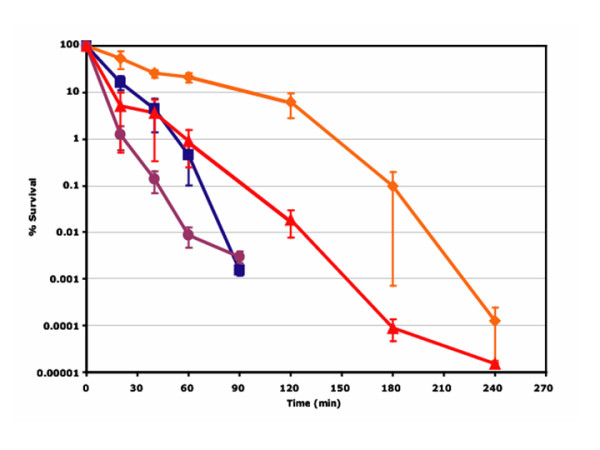
Background: Acid tolerance in Escherichia coli O157:H7 contributes to persistence in its bovine host and is thought to promote passage through the gastric barrier of humans. Dps (DNA-binding protein in starved cells) mutants of E. coli have reduced acid tolerance when compared to the parent strain although the role of Dps in acid tolerance is unclear. This study investigated the mechanism by which Dps contributes to acid tolerance in E. coli O157:H7.
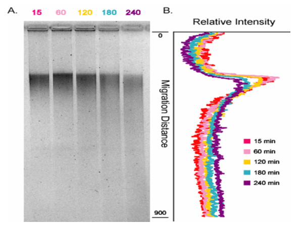
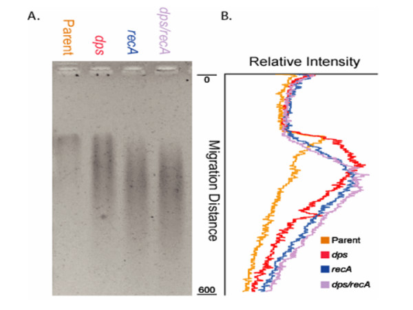
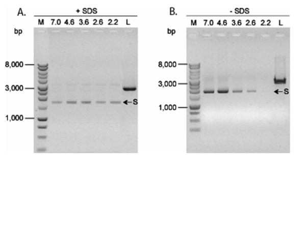
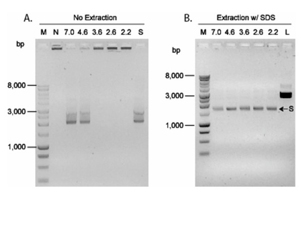
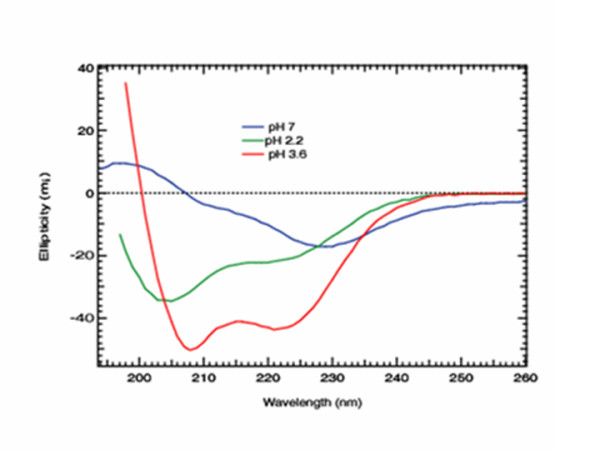
Results: The results from this study showed that acid stress lead to damage of chromosomal DNA, which was accentuated in dps and recA mutants. The use of Bal31, which cleaves DNA at nicks and single-stranded regions, to analyze chromosomal DNA extracted from cells challenged at pH 2.0 provided in vivo evidence of acid damage to DNA. The DNA damage in a recA mutant further corroborated the hypothesis that acid stress leads to DNA strand breaks. Under in vitro assay conditions, Dps was shown to bind plasmid DNA directly and protect it from acid-induced strand breaks. Furthermore, the extraction of DNA from Dps-DNA complexes required a denaturing agent at low pH (2.2 and 3.6) but not at higher pH (>pH4.6). Low pH also restored the DNA-binding activity of heat-denatured Dps. Circular dichroism spectra revealed that at pH 3.6 and pH 2.2 Dps maintains or forms alpha-helices that are important for Dps-DNA complex formation.
Conclusion: Results from the present work showed that acid stress results in DNA damage that is more pronounced in dps and recA mutants. The contribution of RecA to acid tolerance indicated that DNA repair was important even when Dps was present. Dps protected DNA from acid damage by binding to DNA. Low pH appeared to strengthen the Dps-DNA association and the secondary structure of Dps retained or formed alpha-helices at low pH. Further investigation into the precise interplay between DNA protection and damage repair pathways during acid stress are underway to gain additional insight.
Figures

Similar articles
-
Chromosomal fragmentation in dUTPase-deficient mutants of Escherichia coli and its recombinational repair.Mol Microbiol. 2004 Mar;51(5):1279-95. doi: 10.1111/j.1365-2958.2003.03924.x. Mol Microbiol. 2004. PMID: 14982624
-
RecA433 cells are defective in recF-mediated processing of disrupted replication forks but retain recBCD-mediated functions.Mutat Res. 2008 Oct 14;645(1-2):19-26. doi: 10.1016/j.mrfmmm.2008.08.002. Epub 2008 Aug 20. Mutat Res. 2008. PMID: 18782580
-
H-NS controls metabolism and stress tolerance in Escherichia coli O157:H7 that influence mouse passage.BMC Microbiol. 2006 Aug 15;6:72. doi: 10.1186/1471-2180-6-72. BMC Microbiol. 2006. PMID: 16911800 Free PMC article.
-
Recombinational DNA repair: the ignored repair systems.Bioessays. 2004 Dec;26(12):1322-6. doi: 10.1002/bies.20109. Bioessays. 2004. PMID: 15551273 Review.
-
Structure, function and regulation of the DNA-binding protein Dps and its role in acid and oxidative stress resistance in Escherichia coli: a review.J Appl Microbiol. 2011 Feb;110(2):375-86. doi: 10.1111/j.1365-2672.2010.04890.x. Epub 2010 Dec 8. J Appl Microbiol. 2011. PMID: 21143355 Review.
Cited by
-
Dps-like proteins: structural and functional insights into a versatile protein family.Cell Mol Life Sci. 2010 Feb;67(3):341-51. doi: 10.1007/s00018-009-0168-2. Epub 2009 Oct 14. Cell Mol Life Sci. 2010. PMID: 19826764 Free PMC article. Review.
-
Coexisting/Coexpressing Genomic Libraries (CoGeL) identify interactions among distantly located genetic loci for developing complex microbial phenotypes.Nucleic Acids Res. 2011 Dec;39(22):e152. doi: 10.1093/nar/gkr817. Epub 2011 Oct 5. Nucleic Acids Res. 2011. PMID: 21976725 Free PMC article.
-
Comparative Genome-Wide Transcriptome Analysis of Brucella suis and Brucella microti Under Acid Stress at pH 4.5: Cold Shock Protein CspA and Dps Are Associated With Acid Resistance of B. microti.Front Microbiol. 2021 Dec 13;12:794535. doi: 10.3389/fmicb.2021.794535. eCollection 2021. Front Microbiol. 2021. PMID: 34966374 Free PMC article.
-
Molecular mechanism of engineered Zymomonas mobilis to furfural and acetic acid stress.Microb Cell Fact. 2023 May 2;22(1):88. doi: 10.1186/s12934-023-02095-1. Microb Cell Fact. 2023. PMID: 37127628 Free PMC article.
-
YybT is a signaling protein that contains a cyclic dinucleotide phosphodiesterase domain and a GGDEF domain with ATPase activity.J Biol Chem. 2010 Jan 1;285(1):473-82. doi: 10.1074/jbc.M109.040238. Epub 2009 Nov 9. J Biol Chem. 2010. PMID: 19901023 Free PMC article.
References
-
- Johnson LR. Gastric Secretion. In: Johnson LR, editor. Gastrointestinal physiology. 5. St. Louis, MO: Mosby; 1997. pp. 69–88.
-
- Furuta GT, Walker WA. Nonimmune defense mechanisms of the gastrointestinal tract. In: Blaser, Martin J, Smith, Phillip D, Ravdin, Jonathan I, Greenberg, Harry B, Guerrant, Richard L, editor. Infections of the gastrointestinal tract. Vol. 1. New York, NY: Raven Press; 1995. pp. 89–105.
-
- Smith JL. The role of gastric acid in preventing foodborne disease and how bacteria overcome acid conditions. J Food Prot. 2003;66:1292–1303. - PubMed
-
- Lehninger AL, Nelson DL, Cox MM. Principles of biochemistry. 2. New York, N.Y.: Worth Publishers; 1993.
Publication types
MeSH terms
Substances
LinkOut - more resources
Full Text Sources
Molecular Biology Databases
