Comparative Study
. 2008 Oct;19(10):1483-9.
doi: 10.1016/j.jvir.2008.06.017.

Comparison of hypoxia-inducible factor-1alpha expression before and after transcatheter arterial embolization in rabbit VX2 liver tumors

Affiliations
Comparative Study

Comparison of hypoxia-inducible factor-1alpha expression before and after transcatheter arterial embolization in rabbit VX2 liver tumors

Sumeet Virmani et al. J Vasc Interv Radiol. 2008 Oct.

Abstract

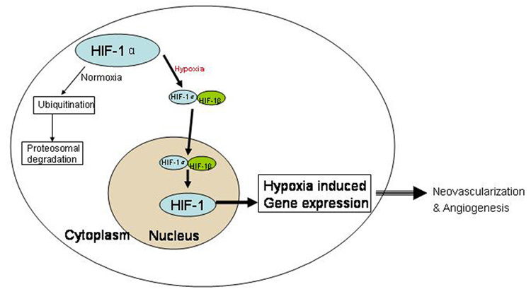
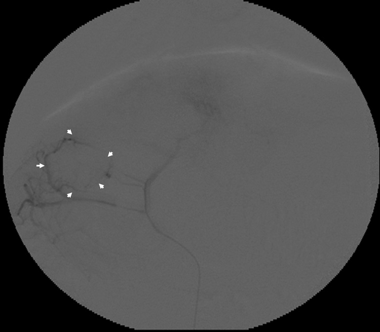
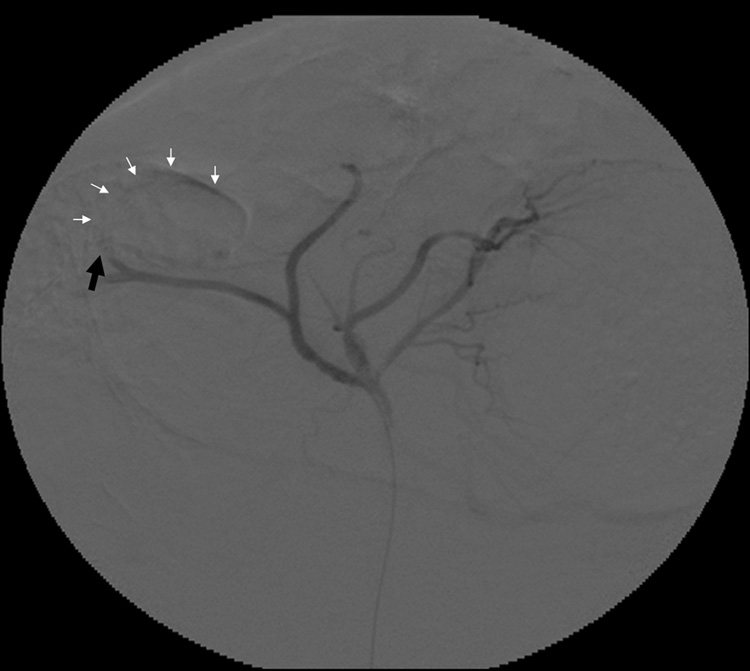
Purpose: To test the hypothesis that transcatheter arterial embolization (TAE) induces expression of hypoxia-inducible factor-1alpha (HIF-1alpha) within the same rabbit VX2 liver tumor.

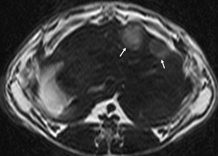
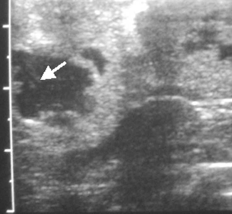
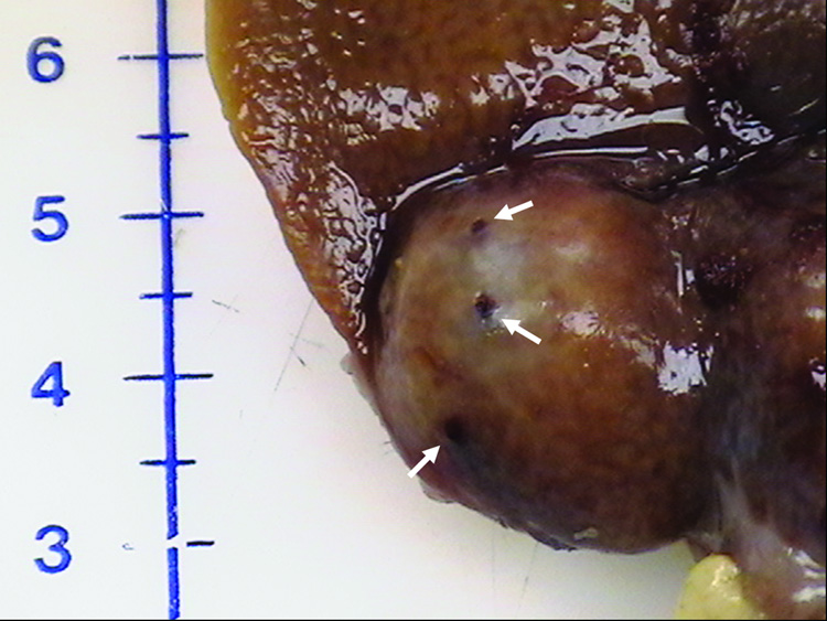
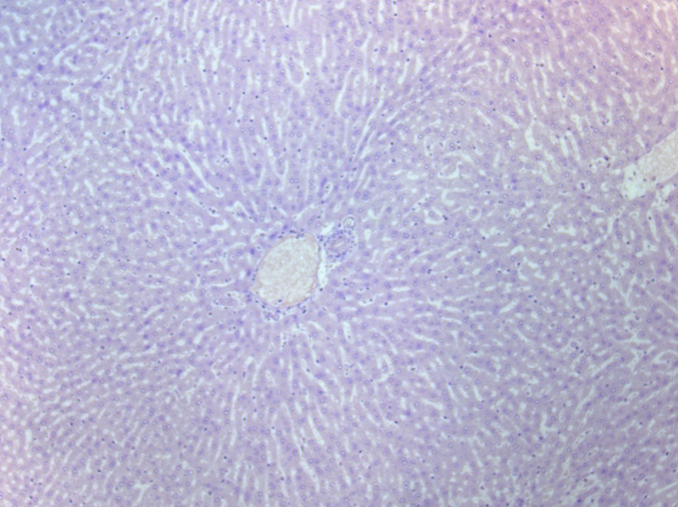
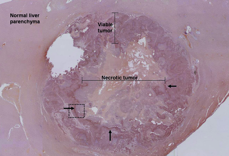
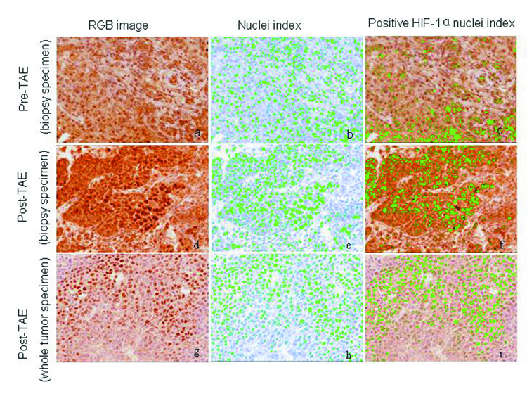
Materials and methods: Seven VX2 tumors were grown in the livers of five New Zealand white rabbits. Ultrasonography-guided biopsy was performed before and 10 minutes after TAE in all tumors. Pre- and post-TAE tumor biopsy specimens along with post-TAE whole liver tumor sections were stained with HIF-1alpha antibody and analyzed for percentage of HIF-1alpha-positive nuclei by using a spectral unmixing system mounted on a high-powered microscope. Statistical data comparisons were performed with the Wilcoxon signed-rank test (alpha = 0.05).

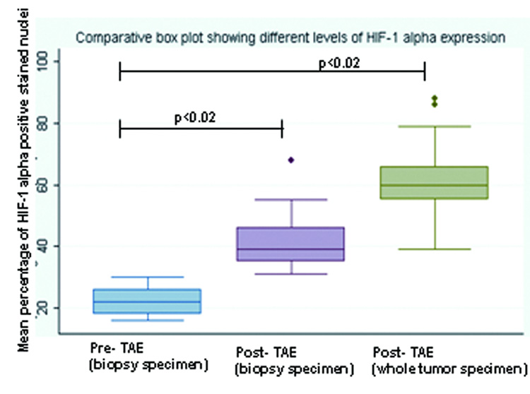
Results: TAE of liver tumors resulted in a statistically significant increase in the mean percentage of HIF-1alpha expression. The mean percentage of HIF-1alpha-positive stained nuclei increased from 23% +/- 3.5 in pre-TAE biopsy specimens to 41% +/- 8.7 in post-TAE biopsy specimens (P < .02). The increase was even more significant when the mean percentage of HIF-1alpha-positive stained nuclei from the same pre-TAE biopsy specimens was compared with sections from post-TAE whole tumor specimens (60% +/- 8.9, P < .02).

Conclusions: The results of this study revealed that hypoxia caused by TAE of VX2 liver tumors activates HIF-1alpha, a transcription factor that in turn regulates other pro-angiogenic factors.


Figures

Figure 1
Figure 1
HIF-1 α activity under normoxic and hypoxic conditions. HIF-1 is a major transcription factor that is composed of two subunits: HIF-1 α and HIF-1 β (beta). Under normoxic conditions, HIF-1 β is constitutively expressed and HIF-1 α is targeted to proteosomal degradation via ubiquitination. On the other hand during hypoxic conditions when oxygen concentration is low, HIF-1 α is stabilized and translocates to nucleus, where it dimerizes with HIF-1 β to form functional HIF-1. This altered redox state occurring in the cells experiencing hypoxia in turn indicates gene transcription of several angiogenic factors.
Figure 2
Figure 2
(a) Axial T2-w MR image showing two VX2 tumor foci in the rabbit’s liver. Tumors show increased signal intensity (arrows) relative to remaining liver; (b) abdominal ultrasound of the rabbit liver showing a hypo-echoic tumor. Also seen is the biopsy needle (arrow) at the center of the tumor.
Figure 2
Figure 2
(a) Axial T2-w MR image showing two VX2 tumor foci in the rabbit’s liver. Tumors show increased signal intensity (arrows) relative to remaining liver; (b) abdominal ultrasound of the rabbit liver showing a hypo-echoic tumor. Also seen is the biopsy needle (arrow) at the center of the tumor.
Figure 3
Figure 3
Representative rabbit hepatic arteriograms before (a) and after (b) embolization of the left hepatic artery, which supplied the targeted VX2 liver tumor. The peripheral portion of the tumor is hypervascular (white arrows) prior to embolization (a). After embolization (b) there is abrupt cut off (black arrow) of the feeding artery without any remaining peripheral hypervascularity.
Figure 3
Figure 3
Representative rabbit hepatic arteriograms before (a) and after (b) embolization of the left hepatic artery, which supplied the targeted VX2 liver tumor. The peripheral portion of the tumor is hypervascular (white arrows) prior to embolization (a). After embolization (b) there is abrupt cut off (black arrow) of the feeding artery without any remaining peripheral hypervascularity.
Figure 4
Figure 4
Harvested rabbit VX2 Liver tumor. Photograph showing multiple biopsy needle puncture sites (arrows) on the tumor surface. No evidence of any significant surrounding hemorrhage can be seen.
Figure 5
Figure 5
Immunohistochemical staining: (a) High-power photomicrograph (original magnification, X 20) of normal liver parenchyma adjacent to VX2 tumor (post-TAE) showing absence of HIF-1 α staining in these cells. (b) High-power photomicrograph (original magnification, X 2.5) of the whole tumor (post- TAE) showing the maximum intensity of HIF-1 α staining at the junction of the necrotic and the viable tumor tissue. Images were stitched together using Canon PhotoStitch 3.1 software. (c) High-power photomicrograph (original magnification, X 10) showing the dark brown nuclear staining, positive for the presence of HIF-1 α staining in these cells.
Figure 5
Figure 5
Immunohistochemical staining: (a) High-power photomicrograph (original magnification, X 20) of normal liver parenchyma adjacent to VX2 tumor (post-TAE) showing absence of HIF-1 α staining in these cells. (b) High-power photomicrograph (original magnification, X 2.5) of the whole tumor (post- TAE) showing the maximum intensity of HIF-1 α staining at the junction of the necrotic and the viable tumor tissue. Images were stitched together using Canon PhotoStitch 3.1 software. (c) High-power photomicrograph (original magnification, X 10) showing the dark brown nuclear staining, positive for the presence of HIF-1 α staining in these cells.
Figure 6
Figure 6
Immunohistochemical staining for HIF-1α. Pre- TAE (biopsy specimen), post- TAE (biopsy specimen) and post- TAE (whole tumor specimen) were analyzed for percentage of HIF-1 α positive nuclei using the Nuance spectral unmixing system. After obtaining the RGB images (images a, d and g), nuclei / HPF were counted (images: b, e and h) using the software (marked green) and then analyzed for nuclei with positive HIF-1α staining (marked green) (images c, f and i). TAE of liver tumors resulted in a statistically significant increase in the mean percentage of HIF-1 α expression.
Figure 7
Figure 7
Box plot showing different levels of HIF-1 α expression in the pre- TAE (biopsy specimen), post- TAE (biopsy specimen) and post- TAE (whole tumor specimen). The boxes represent the inter-quartile range (IQR), the line across the box indicates the median and lines extending from the box represent the 1.5 times the IQR. Values outside the IQR represent the outliers.

References

    1. Llovet JM. Updated treatment approach to hepatocellular carcinoma. J Gastroenterol. 2005;40:225–235. - PubMed
    1. Llovet JM, Schwartz M, Mazzaferro V. Resection and liver transplantation for hepatocellular carcinoma. Semin Liver Dis. 2005;25:181–200. - PubMed
    1. Llovet JM, Bruix J. Systematic review of randomized trials for unresectable hepatocellular carcinoma: Chemoembolization improves survival. Hepatology. 2003;37:429–442. - PubMed
    1. Liao XF, Yi JL, Li XR, Deng W, Yang ZF, Tian G. Angiogenesis in rabbit hepatic tumor after transcatheter arterial embolization. World J Gastroenterol. 2004;10:1885–1889. - PMC - PubMed
    1. Seki T, Tamai T, Ikeda K, et al. Rapid progression of hepatocellular carcinoma after transcatheter arterial chemoembolization and percutaneous radiofrequency ablation in the primary tumour region. Eur J Gastroenterol Hepatol. 2001;13:291–294. - PubMed

Publication types

MeSH terms

Substances