. 2008 Oct 28;105(43):16626-30.
doi: 10.1073/pnas.0808649105. Epub 2008 Oct 15.

Mesothelium contributes to vascular smooth muscle and mesenchyme during lung development

Affiliations

Mesothelium contributes to vascular smooth muscle and mesenchyme during lung development

Jianwen Que et al. Proc Natl Acad Sci U S A. .

Abstract

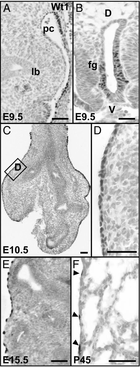
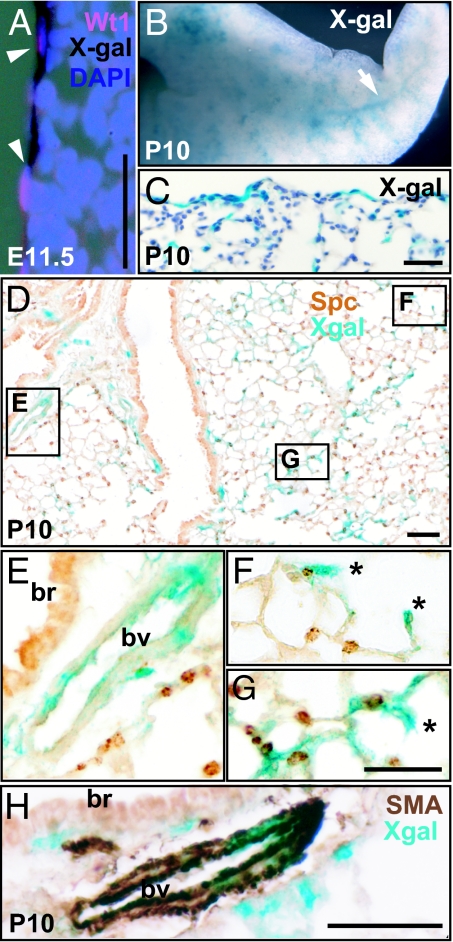
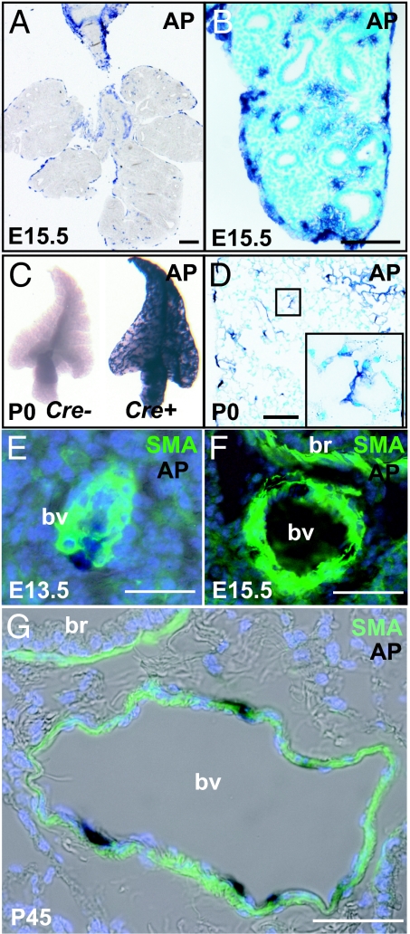
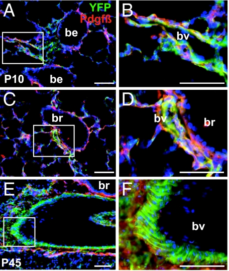
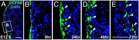
During mouse development, the sophisticated vascular network of the lung is established from embryonic day (E) approximately 10.5 and continues to develop postnatally. This network is composed of endothelial cells enclosed by vascular smooth muscle, pericytes, and other mesenchymal cells. Recent in vivo lineage labeling studies in the developing heart and intestine suggest that some of the vascular smooth muscle cells arise from the surface mesothelium. In the developing lung, the Wilm's tumor 1 gene (Wt1) is expressed only in the mesothelial cells. Therefore, we lineage-labeled the mesothelium in vivo by using a Wt1-Cre transgene in combination with either Rosa26R(lacZ), Rosa26R(CAG-hPLAP), or Rosa26R(EYFP) reporter alleles. In all three cases, cells derived from lineage-labeled mesothelium are found inside the lung and as smooth muscle actin (SMA) and PDGF receptor-beta positive cells in the walls of pulmonary blood vessels. To corroborate this finding, we used 5-(and-6)-carboxy-2',7'-dichlorofluorescein diacetate, succinimidyl ester "mixed isomers" (CCFSE) dye to label mesothelial cells on the surface of the embryonic lung. Over the course of 72-h culture, dye-labeled cells also appear within the lung mesenchyme. Together, our data provide evidence that mesothelial cells serve as a source of vascular smooth muscle cells in the developing lung and suggest that a conserved mechanism applies to the development of blood vessels in all coelomic organs.


Conflict of interest statement

The authors declare no conflict of interest.

Figures

Fig. 1.
Fig. 1.
Expression of Wt1 protein in the developing and adult lung. (A) At E9.5, Wt1 protein is not detected in the primary lung buds, but in mesothelial cells along the wall of the pericardio-peritoneal cavity. (B) Wt1-positive cells in the ventral epithelium of the unseparated foregut. (C) At E10.5, Wt1-positive mesothelium now covers the surface of both the trachea and lungs. (D) A magnified view of the boxed region in C to show the Wt1-positive and tightly packed mesothelial cells. Wt1-positive mesothelial cells at E15.5 (E) and in the adult (arrowheads) (F). Note that at all times examined no Wt1-positive cells are present either in the epithelium or mesenchyme within the lung; lb, lung bud; pc, pericardio-peritoneal cavity; fg, foregut. (Scale bars, 50 μm.)
Fig. 2.
Fig. 2.
Lineage-labeled mesothelium and its descendents in Wt1-Cre;Rosa26RlacZ lungs. (A) Colocalization of X-Gal staining (black) and Wt1 protein (red) on the surface of E11.5 lung. Nuclei are counterstained with DAPI. (B) Whole mount X-Gal staining of P10 lung. The arrow indicates the network of X-Gal stained cells within the lung. (C and D) Sections after whole-mount X-Gal staining to show that X-Gal positive cells are located both on the surface and within the lung tissue. (D–G) Lung section stained with antibody against SftpC. (E–G) Magnified views of the boxed regions in D. (E) X-Gal positive cells are incorporated into the artery close to the bronchus. (F and G) Two examples of X-Gal cells in alveoli possibly pericyte, endothelial cell, or myofibroblast (indicated by *). (H) Colocalization of alpha-SMA (brown) and X-Gal (blue) in the artery wall next to a bronchus. Note that smooth muscle cells underneath the bronchus do not colocalize with X-Gal; br, bronchus; bv, blood vessel. (Scale bars, 50 μm.)
Fig. 3.
Fig. 3.
Localization of lineage-labeled mesothelial cells and their descendants in Wt1-Cre;Rosa26RCAG-hPLAP lungs. (A and B) Sections of E15.5 lung after whole-mount staining for AP. Note that AP-positive cells are present both on the surface and inside the lung. (C) Whole-mount view of a lobe of AP-stained Wt1-Cre;Rosa26RCAG-hPLAP P0 lung (Left) and Rosa26RCAG-hPLAP control lung, which has no Wt1-Cre transgene (Right). (D) Section of P0 lung after whole-mount AP staining. Inset shows AP-positive cells in the alveoli. (E–G) Colocalization of SMA and AP in some of the vascular smooth muscle cells at E13.5 (E), E15.5 (F), and P45 (G); br, bronchus; bv, blood vessel. [Scale bars: 100 μm (A–D) and 50 μm (E–G).]
Fig. 4.
Fig. 4.
Lineage-labeled cells in Wt1-Cre;Rosa26REYFP lungs. (A–F) Immunohistochemistry of sections of P10 (A–D) and P45 (E–F) lungs from Wt1-Cre;Rosa26REYFP mice with antibody to GFP (green) and PDGF receptor-beta (red). (B, D, and F) Magnified view of the boxed regions in A, C, and E, respectively. Note the presence of lineage-labeled cells that also express PDGF receptor-beta on their surface in the relatively thin walls of vessels closely associated with bronchioles (A–D) and in the thicker walls of a vessel (presumed artery) associated with a larger bronchus (E and F). Note also in F the typical organization of lineage-labeled smooth muscle in the thick mural wall of the presumptive artery. In all sections lineage-labeled cells are absent from the population of airway smooth muscle. All nuclei are counterstained with DAPI; be, bronchiole; br, bronchus; bv, blood vessel. (Scale bars, 50 μm.)
Fig. 5.
Fig. 5.
In vitro culture of embryonic lung with dye-labeled mesothelium. (A–E) Distribution of CCFSE-labeled cells after culturing for indicated periods of time (A, 0 h). (B) A magnified view of the boxed region in A. Note that the majority of the surface mesothelial cells are labeled with dye. (C) After 24 h of culture, some dye-labeled cells are present beneath the surface. After culture for 48 (D) and 72 h (E), some dye-labeled are present within the lung tissue. Arrows indicate dye-labeled cells within the lung tissue. All nuclei are counterstained with DAPI. (Scale bars, 50 μm.)

References

    1. Wang Z, Shu W, Lu MM, Morrisey EE. Wnt7b activates canonical signaling in epithelial and vascular smooth muscle cells through interactions with Fzd1, Fzd10, and LRP5. Mol Cell Biol. 2005;25:5022–5030. - PMC - PubMed
    1. White AC, Lavine KJ, Ornitz DM. FGF9 and SHH regulate mesenchymal Vegfa expression and development of the pulmonary capillary network. Development. 2007;134:3743–3752. - PMC - PubMed
    1. deMello DE, Reid LM. Embryonic and early fetal development of human lung vasculature and its functional implications. Pediatr Dev Pathol. 2000;3:439–449. - PubMed
    1. Perez-Pomares JM, et al. Origin of coronary endothelial cells from epicardial mesothelium in avian embryos. Int J Dev Biol. 2002;46:1005–1013. - PubMed
    1. Zhou B, et al. Epicardial progenitors contribute to the cardiomyocyte lineage in the developing heart. Nature. 2008;454:109–113. - PMC - PubMed

Publication types

Substances

LinkOut - more resources