. 2008 Oct 28;105(43):16707-12.
doi: 10.1073/pnas.0808488105. Epub 2008 Oct 15.

Striatal progenitors derived from human ES cells mature into DARPP32 neurons in vitro and in quinolinic acid-lesioned rats

Affiliations

Striatal progenitors derived from human ES cells mature into DARPP32 neurons in vitro and in quinolinic acid-lesioned rats

Laetitia Aubry et al. Proc Natl Acad Sci U S A. .

Abstract

Substitutive cell therapy using fetal striatal grafts has demonstrated preliminary clinical success in patients with Huntington's disease, but the logistics required for accessing fetal cells preclude its extension to the relevant population of patients. Human embryonic stem (hES) cells theoretically meet this challenge, because they can be expanded indefinitely and differentiated into any cell type. We have designed an in vitro protocol combining substrates, media, and cytokines to push hES cells along the neural lineage, up to postmitotic neurons expressing striatal markers. The therapeutic potential of such hES-derived cells was further substantiated by their in vivo differentiation into striatal neurons following xenotransplantation into adult rats. Our results open the way toward hES cell therapy for Huntington's disease. Long-term proliferation of human neural progenitors leads, however, to xenograft overgrowth in the rat brain, suggesting that the path to the clinic requires a way to switch them off after grafting.


Conflict of interest statement

The authors declare no conflict of interest.

Figures

Fig. 1.
Fig. 1.
Phenotypic characterization of striatal progenitors and neurons generated in vitro from hES cells. (A) Outline of the multistep protocol for the differentiation of striatal progenitors and neurons. DIV, days in vitro. (B and C) Proliferative neural rosette cells coexpress Ki67 (green), nestin (red), and Pax6 (red). Few neural cells matured into MAP2+ (green) neurons. (D–G) Neurons at stage E (MAP2+ or TuJ1+; green) expressing striatal markers DARPP32, GABA, calretinin (CalR), and calbindin (CalB) (red). (H) Proportions of cells immunopositive for Oct4 (stage D), nestin, Pax6, Tuj1, MAP2, or GFAP at stages D and E. (I) Proportion of MAP2-positve neurons expressing DARPP32, GABA, CalB, CalR, and TH at stage E. (Scale bar: 100 μm.) ***, P < 0.001 (n = 5).
Fig. 2.
Fig. 2.
Expression of key regulatory genes during differentiation. (A and B) Q-PCR analyses of undifferentiated hES cells, stages A, B, D, E, and human FB. (A) Time-dependent gene expression suggesting the sequential differentiation of hES cells (NANOG+) into neuroectodermal cells (SIX3+), neural precursors (SOX1+), and neurons (MAP2+). (B) Sequential gene expression of markers of developing forebrain (BF1), ganglionic eminences (GSH2 and DLX2), and adult striatum (DARPP32, GAD67, and CALB) suggesting a enrichment in striatal progenitors. (C) Striatal (GSH2 and DLX2) and telencephalic gene (BF1) expression in stage C culture upon exposure for 12 days (stages A–C) to BDNF (−; white bars) or BDNF, SHH, and DKK1 (+; black bars) showing the regulation of the striatal markers by extrinsic factors.*, P < 0.05; **, P < 0.01; ***, P < 0.001 (n = 3 for A and B and n = 5 for C).
Fig. 3.
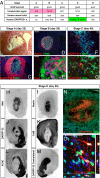
Fig. 3.
Comparative in vivo assessment of different cell preparations 4–6 weeks after xenografting in rat. (A) Key properties of the grafted cells (stages A to E) after transplantation. (B and C) Formation of nonneuroectodermal teratoma-like regions after transplantation of cells at stage A. (D and E) Cells grafted (HNA+; green) at stage B express the neural markers nestin (red), Pax6 (red), and NCAM (green) and are organized in rosette structures in vivo. (F and G) Few human HNA+ (green)/DARPP32+ (red) neurons derived in vivo from stage C. (H–O) In vivo phenotypic characterization of grafts transplanted at stage D. Stage D grafts are HNA+ (H) and nestin+ (I) and contain areas where DARPP32+ (red) cells are clustered (J and N) and have a neuronal morphology as shown in O in a confocal reconstruction; in the same areas, cells may express calbindin (red) (K) but not AChE (L). (M) Contralateral section labeled for DARPP32 shown as a control. (Scale bars: B–D, 1 mm; E–G, N, and O, 100 μm.)
Fig. 4.
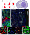
Fig. 4.
Grafted HNA+ cells (stage D, DIV 45) matured into DARPP32 neurons 3–5 months after transplantation. (A–C) DARPP32+ (green)/HNA+ (red) cells are (A and B) spread over the graft and display a neuronal morphology, as shown in C in a confocal reconstruction. (D and E) DARPP32+ (green) cells coexpressed the neuronal marker MAP2 (red). (F) Location of images A, B, and D on a schematic coronal section outline showing the graft location (red) and DARPP32+ host region (green). (Scale bars: A, 200 μm; B and D, 100 μm; C and E, 50 μm.)
Fig. 5.
Fig. 5.
Massive proliferation of neural progenitors 3–5 months after transplantation. (A) Cresyl violet-stained coronal section (right) and outlines of serial coronal sections spanning over ≈10 mm (left) illustrating the size of the graft (red) throughout the host brain. (B–D) Proliferative (Ki67+; red) neural regions (Pax6+; green; or human-specific nestin+; green) are spread all over the graft. (E and F) Expanded and differentiated cells derived from 3-month-old graft dissection. (E–H) Graft-derived neural cultures (nestin+; red) can generate neurons (MAP2+; green) (F) and GFAP+ (red) glial cells (G). Only occasional DARPP32+/MAP2+ neurons (yellow) are generated. (Scale bars: B, 200 μm; C and D, 1 mm; E–H, 100 μm.)

References

    1. Peschanski M, Bachoud-Levi AC, Hantraye P. Integrating fetal neural transplants into a therapeutic strategy: The example of Huntington's disease. Brain. 2004;127:1219–1228. - PubMed
    1. Bachoud-Levi AC, et al. Effect of fetal neural transplants in patients with Huntington's disease 6 years after surgery: A long-term follow-up study. Lancet Neurol. 2006;5:303–309. - PubMed
    1. Bachoud-Levi AC, et al. Motor and cognitive improvements in patients with Huntington's disease after neural transplantation. Lancet. 2000;356:1975–1979. - PubMed
    1. Gaura V, et al. Striatal neural grafting improves cortical metabolism in Huntington's disease patients. Brain. 2004;127:65–72. - PubMed
    1. Peschanski M, Dunnett SB. Cell therapy for Huntington's disease, the next step forward. Lancet Neurol. 2002;1:81. - PubMed

Publication types

LinkOut - more resources