. 2008 Dec 5;283(49):34457-68.
doi: 10.1074/jbc.M801465200. Epub 2008 Oct 15.

Interaction of the RAGE cytoplasmic domain with diaphanous-1 is required for ligand-stimulated cellular migration through activation of Rac1 and Cdc42

Affiliations

Interaction of the RAGE cytoplasmic domain with diaphanous-1 is required for ligand-stimulated cellular migration through activation of Rac1 and Cdc42

Barry I Hudson et al. J Biol Chem. .

Abstract

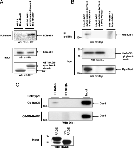
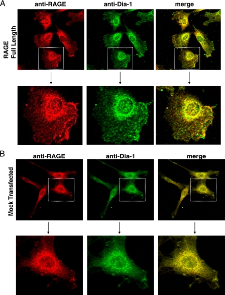
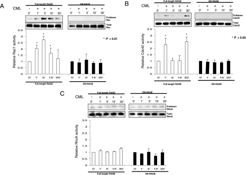
Cellular migration is a fundamental process linked to diverse pathological states such as diabetes and its complications, atherosclerosis, inflammation, and cancer. The receptor for advanced glycation end products (RAGE) is a multiligand cell surface macromolecule which binds distinct ligands that accumulate in these settings. RAGE-ligand interaction evokes central changes in key biological properties of cells, including proliferation, generation of inflammatory mediators, and migration. Although RAGE-dependent signal transduction is critically dependent on its short cytoplasmic domain, to date the proximate mechanism by which this RAGE domain engages and stimulates cytoplasmic signaling pathways has yet to be identified. Here we show that the RAGE cytoplasmic domain interacts with Diaphanous-1 (Dia-1) both in vitro and in vivo. We employed the human RAGE cytoplasmic domain as "bait" in the yeast two-hybrid assay and identified the formin homology (FH1) domain of Dia-1 as a potential binding partner of this RAGE domain. Immunoprecipitation studies revealed that the RAGE cytoplasmic domain interacts with the FH1 domain of Dia-1. Down-regulation of Dia-1 expression by RNA interference blocks RAGE-mediated activation of Rac-1 and Cdc42 and, in parallel, RAGE ligand-stimulated cellular migration. Taken together, these findings indicate that the interaction of the RAGE cytoplasmic domain with Dia-1 is required to transduce extracellular environmental cues evoked by binding of RAGE ligands to their cell surface receptor, a chief consequence of which is Rac-1 and Cdc42 activation and cellular migration. Because RAGE and Dia-1 are implicated in the regulation of inflammatory, vascular, and transformed cell migration, these findings highlight this interaction as a novel target for therapeutic intervention in inflammation, atherosclerosis, diabetes, and cancer.


Figures

FIGURE 1.
FIGURE 1.
Diaphanous-1 is a binding partner for the RAGE cytoplasmic domain. A, schematic of the hDia-1, as adapted from Krebs et al. (17) and Otomo et al. (36). Domains of hDia-1 in the schematic include the Rho GTPase binding domain (Rho-BD), diaphanous inhibitory domain (DID), dimerization domain (DD), coiled-coil region (CC), formin-homology region 1, 2, and 3 (FH1, -2, -3) and diaphanous autoinhibitory domain (DAD). The arrow from 607 to 835 represents the fragment of hDia-1 isolated from a lung cDNA library using the RAGE cytoplasmic domain as bait. The red box represents the novel FH1 sequence identified. B, protein sequence alignment of the isolated yeast two-hybrid clone of hDia-1 (hDia-YIIH). The alignment includes the GenBank™ accession number AF051782 (21) and the full-length sequence of human Dia-1 generated in this study (hDia-Full). The canonical and novel protein sequence differences of hDia-1 identified in this study are indicated in red.
FIGURE 2.
FIGURE 2.
Diaphanous-1 interacts with the RAGE cytoplasmic domain: in vitro and in vivo studies. A, the specificity of the interaction of Dia-1 with the RAGE cytoplasmic domain was analyzed in vitro by GST pulldown assays. GST only or GST-RAGE cytoplasmic tail fusion proteins were incubated with in vitro transcribed/translated His-tagged Dia-YIIH or His tag only (empty vector) labeled with biotinylated lysine-tRNA complex (Transcend™ tRNA). GST pulldown assays were performed, and eluted proteins were analyzed by Western blotting (WB) with streptavidin-horseradish peroxidase (Strep-HRP). An equivalent amount of the GST fusion proteins along with a sample from the in vitro transcription/translation reaction were analyzed by Western blotting using anti-His and anti-GST antibodies (lower panel). B, the RAGE cytoplasmic domain and Dia-1 interaction was analyzed by coimmunoprecipitation (IP) experiments with cell lysates from SK-BR-3 cells transiently transfected with His-tagged RAGE cytoplasmic domain or empty vector and Myc-tagged Dia-1 or empty vector. Cell extracts were immunoprecipitated with anti-His or anti-Myc antibodies and analyzed by Western blotting using anti-His and anti-Myc antibodies. C, C6 glioma cells stably expressing RAGE or the cytoplasmic domain-deleted RAGE (DN-RAGE) were analyzed for interaction with endogenous Dia-1 by coimmunoprecipitation experiments. Cells were serum-starved for 24 h followed by stimulation with CML-HSA (10 μg/ml) for 30 min. Cell extracts were then immunoprecipitated with anti-RAGE antibodies and analyzed by Western blotting with anti-Dia-1 antibodies. Total extract used for immunoprecipitation is indicated as input lysate and was analyzed by Western blotting with anti-RAGE and anti-Dia-1 antibodies. All figures (A-C) are representative images of multiple independent experiments.
FIGURE 3.
FIGURE 3.
RAGE and Dia-1 colocalization in cells. Confocal microscopy was performed using the C6 cells stably transfected with either full-length RAGE (A) or empty vector (Mock) (B) constructs. Cells were treated with RAGE ligand for 30 min, fixed, and immunostained with antibodies to RAGE and Dia-1 followed by respective biotinylated secondary antibodies and streptavidin-linked Alexa-546 (anti-RAGE) and 488 (anti-Dia-1). Higher magnification images of the cells within the stippled boxes in the upper panels are illustrated in the lower panels.
FIGURE 4.
FIGURE 4.
The FH1 domain of hDia-1 is critical for binding to the RAGE cytoplasmic domain. A, schematic diagram of Myc-tagged hDia-1 and truncation mutants used in this study. Numbers indicate amino acid positions in human Dia-1. Rho-BD, Rho GTPase binding domain; DID, diaphanous inhibitory domain; DD, dimerization domain; CC, coiled-coil region; FH1 and -2, formin-homology region 1 and 2; DAD, diaphanous autoinhibitory domain. B, to analyze the domain(s) of Dia-1, which interacts with the RAGE cytoplasmic domain, coimmunoprecipitation (IP) experiments were performed. SK-BR-3 cells were transiently cotransfected with the indicated Myc-tagged hDia-1 constructs and the His-tagged RAGE cytoplasmic domain. Immunoprecipitation was performed with anti-His antibodies followed by Western blotting (WB) with anti-Myc antibodies (left panel). Western blot analysis was performed on input lysate as indicated with anti-Myc and anti-His antibodies for hDia-1 and RAGE cytoplasmic domain constructs, respectively (right panel). Note that the hDia-RhoBD does not bind to the RAGE cytoplasmic domain. All panels (A-B) are representative images of multiple independent experiments.
FIGURE 5.
FIGURE 5.
The RAGE cytoplasmic domain is required for cellular migration. Migration assays were performed with C6 glioma cells stably expressing RAGE or the cytoplasmic domain deleted RAGE (DN-RAGE) to verify the requirement of the RAGE cytoplasmic domain for RAGE-ligand-driven cellular migration. Cells (1 × 10⁵) were seeded into the top of transwell migration chambers and allowed to migrate for 5 h toward RAGE-ligand or media alone negative control placed in the lower chambers. Cells were fixed and stained using cell stain solution, and three independent fields were counted. Results are expressed as the mean ± S.D. (n = 3).
FIGURE 6.
FIGURE 6.
The RAGE cytoplasmic domain is required for activation of Rac-1 and Cdc42 but not RhoA. GTPase activation assays were performed with C6 glioma cells stably expressing RAGE or the cytoplasmic domain deleted RAGE (DN-RAGE), starved for 24 h, treated with CML-HSA (10 μg/ml), and lysed at the indicated times. GTP activated Rac1 (A), Cdc42 (B), and RhoA (C) were subjected to pulldown assays and examined by Western blotting compared with total GTPase in lysate. Results are expressed as the mean ± S.D. (n = 3). Western blots are representative of three independent experiments.
FIGURE 7.
FIGURE 7.
The RAGE cytoplasmic domain is required for Rac-1- and Cdc42-but not RhoA-mediated cellular migration. Migration assays were performed with C6 glioma cells stably expressing RAGE, cotransfected with GFP and DN-Rac1, DN-Cdc42, DN-RhoA, or empty vector. Migration assays were performed as described above, and results are the means ± S.D. (n = 3).
FIGURE 8.
FIGURE 8.
Dia-1 siRNA blocks RAGE ligand-stimulated cellular migration. C6 glioma cells stably expressing RAGE were transfected with siRNAs against Dia-1 or siRNA control. 48 h after transfection cells were lysed, and Dia-1 levels were assessed by quantitative-PCR (A) or Western blotting (WB) using anti-Dia-1 antibodies (B). C, cells were transfected with either Dia-1 siRNA/GFP and scramble siRNA/GFP and, after 48 h, fixed and immunostained with antibodies to Dia-1 followed by respective biotinylated secondary antibodies and streptavidin-linked Alexa-546 (Dia-1). D, cells were cotransfected with GFP and Dia-1/control siRNA, and migration stimulated by CML-HAS or 10% fetal bovine serum control (FBS) was performed and quantified as described above. The results are the means ± S.D. (n = 3).
FIGURE 9.
FIGURE 9.
Dia-1 siRNA blocks RAGE ligand-mediated activation of Rac1 and Cdc42. C6 glioma cells stably expressing RAGE were transfected with siRNAs against Dia-1 or siRNA control. Cells were starved for 24 h, treated with CML-HSA (10 μg/ml), and lysed at the indicated times as established in Fig. 7. GTP-activated Rac1 (A) or Cdc42 (B) was subjected to pulldown assays and examined by Western blotting compared with total GTPase in lysate. Results are expressed as the mean ± S.D. (n = 3). C, Dia-1 knockdown was confirmed in each experiment by Western blot. Importantly, Dia-1 knockdown did not affect RAGE expression. In all cases equal protein loading was demonstrated by glyceraldehyde-3-phosphate dehydrogenase (GAPDH) Western blots (WB). Western blots are representative of three independent experiments.

References

    1. Neeper, M., Schmidt, A. M., Brett, J., Yan, S. D., Wang, F., Pan, Y. C. P., Elliston, K., Stern, D., and Shaw, A. (1992) J. Biol. Chem. 267 14998-15004 - PubMed
    1. Hofmann, M. A., Drury, S., Fu, C. F., Qu, W., Taguchi, A., Lu, Y., Avila, C., Kambham, N., Bierhaus, A., Nawroth, P., Neurath, M. F., Slattery, T., Beach, D., McClary, J., Nagashima, M., Morser, J., Stern, D., and Schmidt, A. M. (1999) Cell 97 889-901 - PubMed
    1. Hori, O., Brett, J., Slattery, T., Cao, R., Zhang, J., Chen, J. X., Nagashima, M., Lundh, E. R., Vijay, S., Nitecki, D., Morser, J., Stern, D., and Schmidt, A. M. (1995) J. Biol. Chem. 270 25752-25761 - PubMed
    1. Yan, S. D., Chen, X., Fu, J., Chen, M., Zhu, H., Roher, A., Slattery, T., Zhao, L., Nagashima, M., Morser, J., Migheli, A., Nawroth, P., Stern, D., and Schmidt, A. M. (1996) Nature 382 685-691 - PubMed
    1. Huttunen, H. J., Fages, C., and Rauvala, H. (1999) J. Biol. Chem. 274 19919-19924 - PubMed

Publication types

Associated data