. 2008 Dec 19;283(51):35579-89.
doi: 10.1074/jbc.M805507200. Epub 2008 Oct 14.

Ischemic injury to kidney induces glomerular podocyte effacement and dissociation of slit diaphragm proteins Neph1 and ZO-1

Affiliations

Ischemic injury to kidney induces glomerular podocyte effacement and dissociation of slit diaphragm proteins Neph1 and ZO-1

Mark C Wagner et al. J Biol Chem. .

Abstract

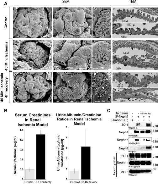
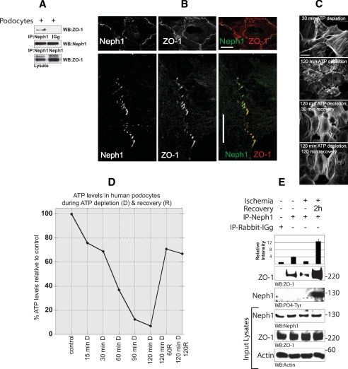
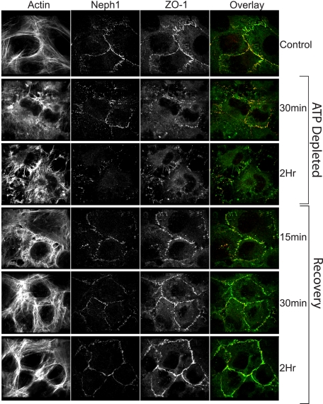
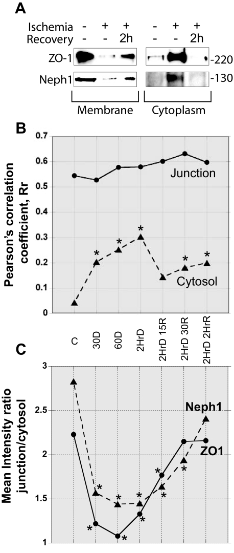
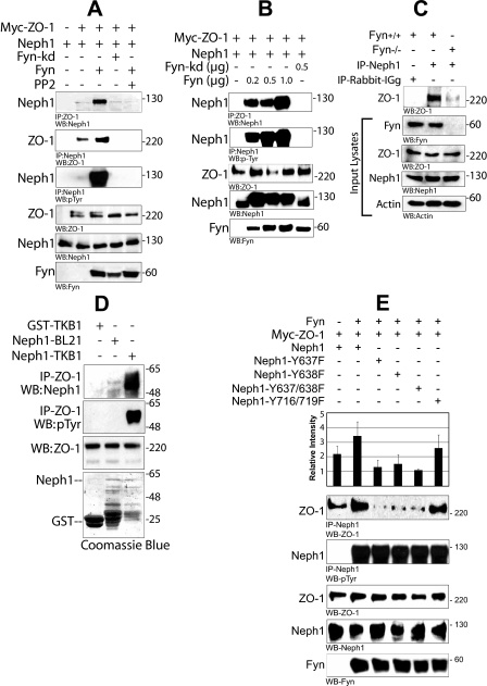
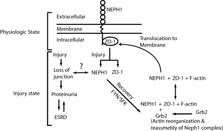
Glomerular injury is often characterized by the effacement of podocytes, loss of slit diaphragms, and proteinuria. Renal ischemia or the loss of blood flow to the kidneys has been widely associated with tubular and endothelial injury but rarely has been shown to induce podocyte damage and disruption of the slit diaphragm. In this study, we have used an in vivo rat ischemic model to demonstrate that renal ischemia induces podocyte effacement with loss of slit diaphragm and proteinuria. Biochemical analysis of the ischemic glomerulus shows that ischemia induces rapid loss of interaction between slit diaphragm junctional proteins Neph1 and ZO-1. To further understand the effect of ischemia on molecular interactions between slit diaphragm proteins, a cell culture model was employed to study the binding between Neph1 and ZO-1. Under physiologic conditions, Neph1 co-localized with ZO-1 at cell-cell contacts in cultured human podocytes. Induction of injury by ATP depletion resulted in rapid loss of Neph1 and ZO-1 binding and redistribution of Neph1 and ZO-1 proteins from cell membrane to the cytoplasm. Recovery resulted in increased Neph1 tyrosine phosphorylation, restoring Neph1 and ZO-1 binding and their localization at the cell membrane. We further demonstrate that tyrosine phosphorylation of Neph1 mediated by Fyn results in significantly increased Neph1 and ZO-1 binding, suggesting a critical role for Neph1 tyrosine phosphorylation in reorganizing the Neph1-ZO-1 complex. This study documents that renal ischemia induces dynamic changes in the molecular interactions between slit diaphragm proteins, leading to podocyte damage and proteinuria.


Figures

FIGURE 1.
FIGURE 1.
Ischemic injury to rat kidney induces podocyte effacement. A, scanning and transmission electron micrographs of kidneys from untreated and ischemic rats at 0 and 4 h after 45 min of ischemia. Foot process structure was lost after ischemia and ischemia, followed by 4 h of recovery. After 45 min of ischemia, glomerular foot processes appear swollen with decreased filtration slits, and by 4 h of recovery, podocyte effacement became prominent with numerous microvilli. SEM, scanning electron micrographs; TEM, transmission electron micrographs; P, podocytes; GBM, glomerular basement membrane; SD, slit diaphragm. B, serum creatinine concentration and urine albumin/creatinine ratios of control and ischemic rats evaluated at 4 h of recovery (p < 0.04) suggest increased proteinuria compared with control. Injury to rat kidney by ischemia induces dissociation of Neph1 from ZO-1 and podocyte effacement. C, rat glomerular lysate was obtained after 45 min of ischemia and ischemia followed by recovery for 4 h. Neph1 was immunoprecipitated (IP) from these and control untreated glomerular lysate using Neph1 antibody and immunoblotted with ZO-1, Neph1, Nephrin, and Tyr(P) antibodies. Equivalent amounts of Neph1, ZO-1, and Nephrin in the tissue lysates were examined using their respective antibodies. This experiment was performed at least five times with similar results.
FIGURE 2.
FIGURE 2.
Neph1 and ZO-1 are localized at the cell-cell junction in cultured podocytes. A, lysate obtained from the cultured human podocyte cell line was immunoprecipitated (IP) with Neph1 antibody and immunoblotted with Neph1 and ZO-1 antibodies. B, localization of Neph1 and ZO-1 in the podocyte cell line was examined by staining the cells with Neph1 and ZO-1 antibodies. Immunofluorescence analysis was done using confocal microscopy. Colocalization of Neph1 and ZO-1 at the membrane junctions appears yellow on merged images. Ischemia results in dissociation of Neph1 and ZO-1. C and D, podocyte cells were subjected to ATP depletion by antimycin A for the indicated times. Following ATP depletion, the medium was replaced with fresh growth medium, and the cells were grown for an additional 30 and 120 min. At each time point, the cells were stained with actin (C), and relative ATP levels were determined using the Promega Enliten ATP assay system (D). E, the cells were subjected to injury by treatment with ischemia for 120 min. Following injury, the medium in the cells was replaced with fresh growth medium, and the cells were grown for an additional 2 h. The cells were lysed and immunoprecipitated with Neph1 antibody. The presence of ZO-1 in the immune complex was examined by immunoblotting with ZO-1 antibody. Relative binding of ZO-1 with Neph1 during ischemia and recovery is also presented in the form of a bar diagram, where data are presented as a mean of three independent experiments.
FIGURE 3.
FIGURE 3.
Ischemia/reperfusion induces a significant shift in the localization of Neph1 and ZO-1. Podocyte cells were grown on coverslips and subjected to ATP depletion for 30 min or 2 h. The recovery followed a 2-h injury and was performed by replacing the medium with fresh growth medium and growing the cells for the indicated times. At each time point, the cells were stained with Neph1 and ZO-1 antibodies to determine the localization of these proteins. Immunofluorescence analysis was performed using confocal microscopy. To determine the extent of Neph1 and ZO-1 co-localization, each of the figures was analyzed using Image J software, as described under “Materials and Methods.”
FIGURE 4.
FIGURE 4.
Analysis of subcellular fractionation and immunofluorescent data supports changes in localization of ZO-1 and Neph1 during ischemia/reperfusion. A, podocyte cells were grown and subjected to ischemia, followed by recovery. The cells were fractionated to isolate cytoplasmic and membrane fractions, and the amounts of Neph1 and ZO-1 in each fraction were analyzed by Western blotting. B, Pearson's correlation coefficient, Rr, was calculated to address the interaction between ZO-1 and Neph1 control (c), ischemic, and recovery time points. Note the increase in Rr in the cytosol during ATP repletion. C, mean integrated intensities of ZO-1 and Neph1 were calculated in the same junction and cytosol regions used to calculate the Rr. The ratio of junction/cytosol for each protein is shown. An unpaired Student's t test compared each time point to control. Experiments were run a minimum of three times, with each data point representing the analysis of more than eight cell fields.
FIGURE 5.
FIGURE 5.
Fyn increases the interaction between Neph1 and ZO-1 by directly phosphorylating Neph1. A, HEK293 cells were transfected with plasmids encoding FLAG-Neph1, Myc-ZO-1, and Fyn or Fyn-kd (catalytically inactive). ZO-1 or Neph1 was immunoprecipitated from the cell lysate, and immune complexes were evaluated for binding of Neph1 to ZO-1 and phosphorylation of Neph1. The experiment was repeated five times with similar results. B, plasmids encoding Myc-ZO-1 and FLAG-Neph1 were co-transfected with either Fyn-kd or increasing amounts of Fyn plasmids in HEK-293 cells. ZO-1 and Neph1 were immunoprecipitated from the cell lysate and immunoblotted with Neph1 and Tyr(P) (phosphotyrosine) antibodies, respectively. C, isolated glomeruli obtained from Fyn null mice or wild type mice (with the same genetic background sv129 strain) were lysed and immunoprecipitated (IP) with Neph1 antibody and immunoblotted with ZO-1. This experiment was repeated twice with similar results. D, phosphorylation of Neph1 increases its interaction with ZO-1. ZO-1 was immunoprecipitated from the HEK293 cell lysate and incubated with either GST alone or phosphorylated GST-Neph1 cytoplasmic domain expressed in tyrosine kinase expressing E. coli TKB1 or E. coli BL21. Binding of Neph1 to ZO-1 was analyzed by immunoblotting with Neph1 and phosphotyrosine antibodies. E, phosphorylation of Neph1 on sites 637 and 638 is required for its increased interaction with ZO-1. Plasmids encoding various indicated Neph1 mutants were co-transfected with Myc-ZO-1 and Fyn in HEK-293 cells. Neph1 was immunoprecipitated from the cell lysate, and the immune complexes were analyzed for the binding of ZO-1 and Neph1 phosphorylation by ZO-1 and Tyr(P) antibodies, respectively. Data from four independent experiments were analyzed, and the results presented are means plus S.E. for these experiments.
FIGURE 6.
FIGURE 6.
A schematic representation of the dynamic interaction between Neph1 and ZO-1. The model summarizes the hypothesis suggesting that under basal conditions, Neph1 and ZO-1 exist as a complex. Injury by ischemia induces dissociation of Neph1 and ZO-1 and their movement to cytoplasm with a corresponding loss of slit diaphragm junction. Recovery events that follow induce Neph1 tyrosine phosphorylation in an Src family kinase-dependent manner and lead to reorganization of Neph1 and ZO-1 complex and subsequent localization of this complex to the cell membrane.

References

    1. Molitoris, B. A., Melnikov, V. Y., Okusa, M. D., and Himmelfarb, J. (2008) Nat. Clin. Pract. Nephrol. 4 154-165 - PubMed
    1. Molitoris, B. A., and Sutton, T. A. (2004) Kidney Int. 66 496-499 - PubMed
    1. Kelly, K. J., and Molitoris, B. A. (2000) Semin Nephrol. 20 4-19 - PubMed
    1. Molitoris, B. A. (2003) J. Am. Soc. Nephrol. 14 265-267 - PubMed
    1. Parikh, C. R., Edelstein, C. L., Devarajan, P., and Cantley, L. (2007) J. Investig. Med. 55 333-340 - PubMed

Publication types

MeSH terms