. 2008 Oct 15;68(20):8286-94.
doi: 10.1158/0008-5472.CAN-07-6316.

Suppression of the negative regulator LRIG1 contributes to ErbB2 overexpression in breast cancer

Affiliations

Suppression of the negative regulator LRIG1 contributes to ErbB2 overexpression in breast cancer

Jamie K Miller et al. Cancer Res. .

Abstract

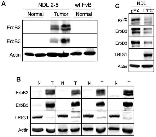
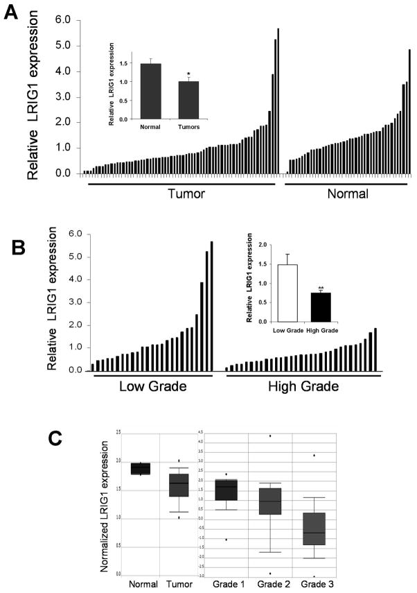
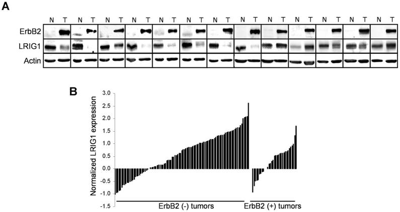
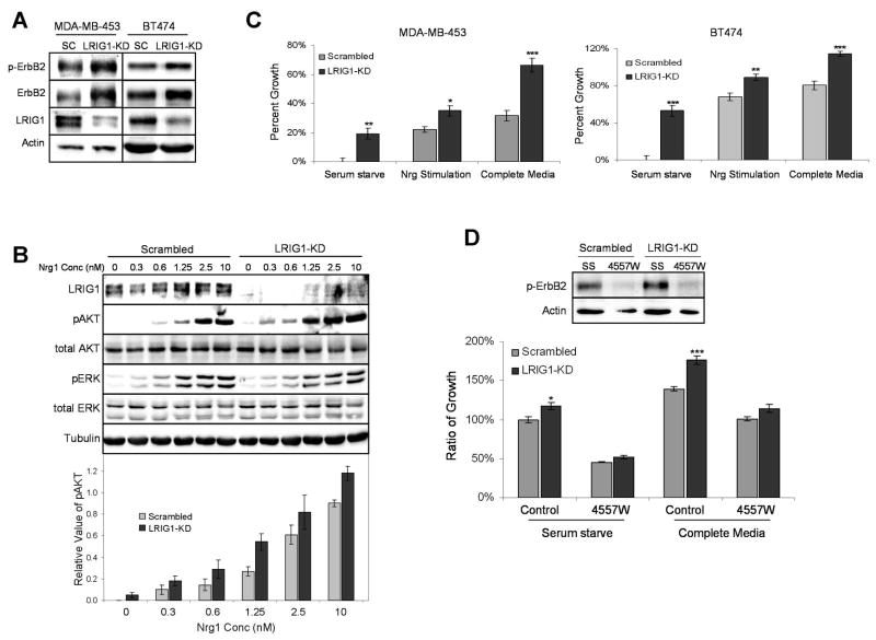
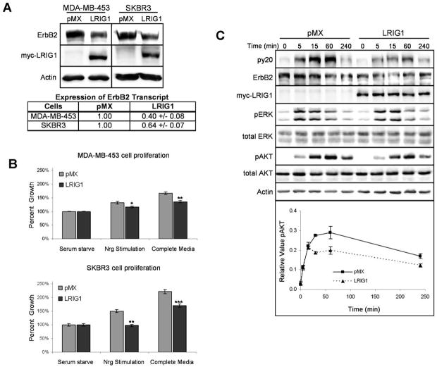
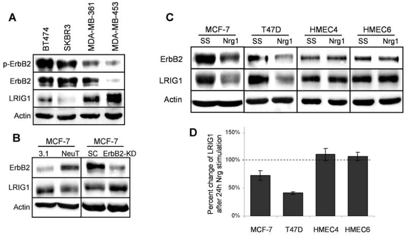
The ErbB2 receptor tyrosine kinase is overexpressed in approximately 25% of breast tumors and contributes to poor patient prognosis and therapeutic resistance. Here, we examine the role of the recently discovered ErbB negative regulator LRIG1 in ErbB2(+) breast cancer. We observe that LRIG1 protein levels are significantly suppressed in ErbB2-induced mammary tumors in transgenic mice as well as in the majority of ErbB2(+) human breast tumors. These observations raise the possibility that LRIG1 loss could contribute to the initiation or growth of ErbB2(+) breast tumors. RNA interference-mediated knockdown of endogenous LRIG1 in the ErbB2-overexpressing breast tumor cell lines MDA-MB-453 and BT474 further elevates ErbB2 in these cells and augments cellular proliferation. In contrast, ectopic expression of LRIG1 reverses these trends. Interestingly, we observe that LRIG1 protein levels are suppressed in response to ErbB receptor activation in breast tumor cells but are unaffected by ErbB activation in immortalized nontransformed breast epithelial cells. Our observations indicate that the suppression of LRIG1 protein levels is a common feature of breast tumors. Moreover, our observations point to the existence of a feed-forward regulatory loop in breast tumor cells where aberrant ErbB2 signaling suppresses LRIG1 protein levels, which in turn contributes to ErbB2 overexpression.


Figures

Figure 1
Figure 1
LRIG1 is suppressed in tumors from MMTV-ErbB2 mice. A, Normal mammary tissue from non-transgenic FvB littermates was collected along with normal and tumor tissue from MMTV-NDL2-5 mice. Lysates from these tissues were blotted for ErbB2, ErbB3, and actin as a loading control. B, Tumor (T) and adjacent normal (N) tissue were collected from the mammary fat pad of NDL2-5 transgenic mice. Lysates from these tissues were blotted for ErbB2, ErbB3, LRIG1, and actin. C, An epithelial cell line produced from a NDL2-5 tumor was transduced with myc-tagged LRIG1 or control vector pMX. Lysates were immunblotted with antibodies to phosphotyrosine (py20), ErbB2, ErbB3, LRIG1, and actin.
Figure 2
Figure 2
LRIG1 levels are decreased in human breast tumors. A, Relative LRIG1 expression was compared in breast tumor and normal specimens via western blot analysis. Each of the 67 tumor and 42 normal samples are plotted in order of increasing LRIG1 levels after normalizing to actin levels. Inset, The mean value of LRIG1 protein expression for each subset is plotted with error bars showing SE. B, Using the Richardson-Bloom grade information available from the pathology reports of 58 of the 67 tumors, relative LRIG1 expression of each tumor is plotted as a function of the tumor grade. Low grade represents tumor grades of 1 and 2, with high grade corresponding to grade 3 tumors. Inset, The average LRIG1 level of each grouping is plotted in the right panel with error bars showing the SE. C, Representative plots of normalized LRIG1 transcript in breast tissue from the Oncomine database are pictured. Each study has a P value of < 0.05.
Figure 3
Figure 3
ErbB2 (+) human tumors display reduced LRIG1 expression. A, Lysates of ErbB2 (+) tumors and patient-matched normal samples were immunoblotted with antibodies to ErbB2, LRIG1, and actin. A pie chart depicting the frequency of LRIG1 loss in these tumors is shown below. B, The Hess study available through Oncomine (27) was queried for transcript expression of LRIG1 in breast tumors and is plotted on the basis of ErbB2 status of the tumors.
Figure 4
Figure 4
Depletion of LRIG1 augments ErbB2 expression and enhances cellular proliferation. A, Lysates from MDA-MB-453 and BT474 cells transfected for 72h with RNAi oligos targeting LRIG1 (LRIG1-KD) or scrambled control (SC) were blotted with antibodies to p-ErbB2, ErbB2, LRIG1, and actin. B, 48h after transfecting with RNAi oligos, MDA-MB-453 cells were serum starved overnight and then treated with increasing concentrations of Nrg1 for 15 min. Whole cell lysates were blotted with antibodies to LRIG1, pAKT, tAKT, pERK, tERK, and tubulin as a loading control. The bottom panel plots the densitometic analysis of the average pAKT signal from three independent experiments for each Nrg1 concentration along with SE. C, 24h after transfecting cells with RNAi oligos as described above, MDA-MB-453 and BT474 cells were placed in serum starve media, in the absence or presence of 2.5 nM Nrg1, or complete media (10% FCS). After 48h, proliferation was measured by an MTT assay. Three independent experiments were conducted with a representative experiment shown. Error bars represent SE of four replicates within this experiment. D, 24h after transfecting cells with RNAi oligos as described above, MDA-MB-453 cells were placed in serum starve or complete media in the absence or presence of the EGFR/ErbB2 inhibitor 4557W. After 48h, proliferation was measured by an MTT assay, with basal proliferation being normalized as 100%. Additionally, whole cell lysates from serum starved cells were collected following treatment and blotted for p-ErbB2 and actin (top panel) to verify inhibition of ErbB2 by 4557W.
Figure 5
Figure 5
Ectopic expression of LRIG1 inhibits breast cancer cell proliferation. A, MDA-MB-453 and SKBR-3 cells were transduced with myc-tagged LRIG1 retrovirus or pMX control. Whole cell lysates were immunoblotted with antibodies for ErbB2, myc, and actin. ErbB2 transcript was measured by real-time PCR analysis. pMX transduced cells were normalized to 1.0 B, MDA-MB-453 and SKBR3 cells transduced with LRIG1 or control pMX vector were treated with serum starve media in the presence or absence of 2.5 nM Nrg1, or with complete media, for 48h. Proliferation was then measured via an MTT assay. Each experiment was repeated in triplicate with a representative experiment shown. Error bars represent SE of four replicates. C, MDA-MB-453 cells transduced with LRIG1 or control pMX vector were stimulated with 1.25 nM Nrg1 for various timepoints. Whole cell lysates were then immunoblotted with antibodies to phosphotyrosine (py20), ErbB2, myc, pAKT, tAKT, pERK, tERK, and actin. Densitometric analysis of the pAKT signal normalized to tAKT is plotted for each timepoint along with SE from three independent experiments.
Figure 6
Figure 6
ErbB2-activation reduces LRIG1 expression in breast tumor cells. A, Whole cell lysates collected from BT474, SKBR3, MDA-MB-361 and MDA-MB-453 cells were blotted with antibodies to p-ErbB2, ErbB2, LRIG1, and actin. B, MCF-7 cells were transiently transfected for 48h with NeuT or control vector pcDNA3.1 (3.1) and cell lysates were blotted for ErbB2, LRIG1, and actin. Additionally, MCF-7 cells were treated with 100 nM scrambled control (SC) or ErbB2 RNAi oligos (ErbB2-KD) for 48h. Lysates were collected and blotted for ErbB2, LRIG1, and actin. C, MCF-7, T47D, HMEC4, and HMEC6 cells were treated with SS media in the absence or presence of 10 nM Nrg1. After 24h, whole cell lysates were collected and samples were immunoblotted with antibodies to ErbB2, LRIG1, and actin. D, Densitometric analysis of LRIG1 protein expression from each cell line in C is plotted along with SE from at least four independent experiments.

References

    1. Sweeney C, Miller JK, Shattuck DL, Carraway KL., 3rd ErbB receptor negative regulatory mechanisms: implications in cancer. J Mammary Gland Biol Neoplasia. 2006;11:89–99. - PubMed
    1. Peschard P, Park M. Escape from Cbl-mediated downregulation: a recurrent theme for oncogenic deregulation of receptor tyrosine kinases. Cancer Cell. 2003;3:519–23. - PubMed
    1. Moasser MM. Targeting the function of the HER2 oncogene in human cancer therapeutics. Oncogene. 2007;26:6577–92. - PMC - PubMed
    1. Holbro T, Beerli RR, Maurer F, Koziczak M, Barbas CF, 3rd, Hynes NE. The ErbB2/ErbB3 heterodimer functions as an oncogenic unit: ErbB2 requires ErbB3 to drive breast tumor cell proliferation. Proc Natl Acad Sci U S A. 2003;100:8933–8. - PMC - PubMed
    1. Naidu R, Yadav M, Nair S, Kutty MK. Expression of c-erbB3 protein in primary breast carcinomas. Br J Cancer. 1998;78:1385–90. - PMC - PubMed

Publication types