. 2008 Oct 15;68(20):8361-8.
doi: 10.1158/0008-5472.CAN-08-0878.

Dual inhibition of PI3Kalpha and mTOR as an alternative treatment for Kaposi's sarcoma

Affiliations

Dual inhibition of PI3Kalpha and mTOR as an alternative treatment for Kaposi's sarcoma

Risa Chaisuparat et al. Cancer Res. .

Abstract

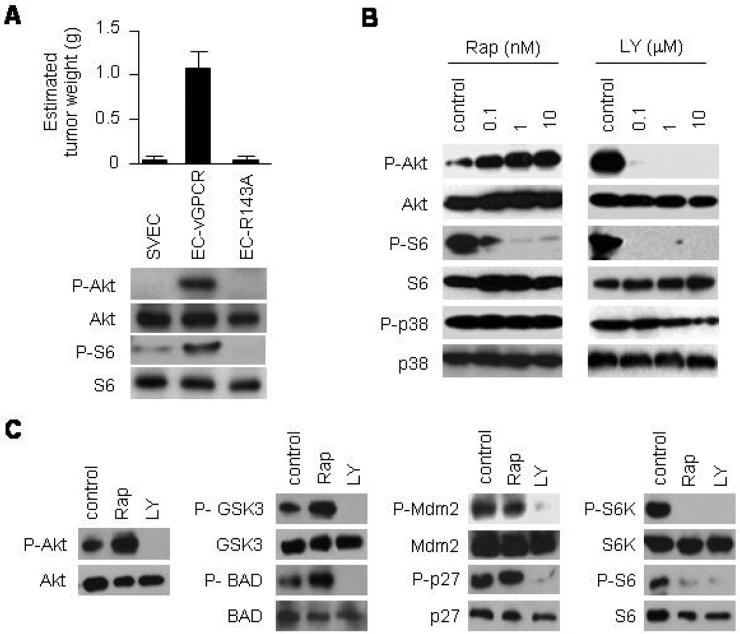
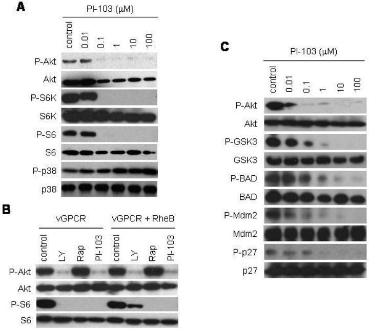
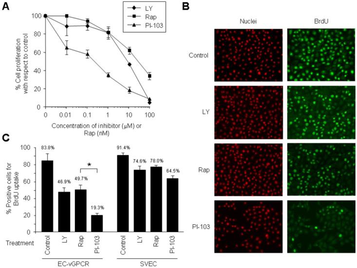
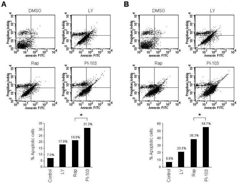
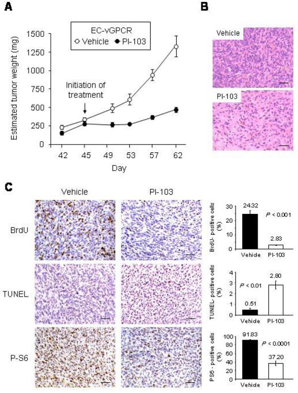
Rapamycin (or sirolimus), the prototypical inhibitor of the mammalian target of rapamycin (mTOR) and an immunosuppressant used for the prevention of renal transplant rejection, has recently emerged as an effective treatment for Kaposi's sarcoma (KS), an enigmatic vascular tumor and a model for pathologic angiogenesis. Indeed, recent work supports a role for mTOR as a central player in the transformation of endothelial cells by the KS-associated herpesvirus-encoded G protein-coupled receptor (vGPCR), the viral oncogene believed to be responsible for causing KS. However, emerging evidence that rapamycin may transiently promote the activation of Akt may limit its use as an anti-KS therapy. Here, we show that activation of Akt in endothelial cells expressing vGPCR is augmented by treatment with rapamycin, resulting in the up-regulation of several Akt proliferative and survival pathways. However, use of a novel dual phosphatidylinositol 3-kinase alpha (PI3Kalpha)/mTOR inhibitor, PI-103, effectively and independently blocked activation of both PI3K and mTOR in vGPCR-expressing endothelial cells. This resulted in more effective inhibition of endothelial cell proliferation and survival in vitro and tumor growth in vivo. Our results suggest that PI-103 may be an effective therapeutic option for the treatment of patients with KS. Moreover, as KS may serve as a model for pathologic angiogenesis, our results further provide the basis for the early assessment of PI-103 as an antiangiogenic chemotherapeutic.


Figures

Figure 1
Figure 1. Rapamycin upregulates Akt signaling in KSHV vGPCR-expressing endothelial cells
(A) Tumor allografts were established in athymic nu/nu mice by injecting immortalized murine endothelial cells (SVECs), or SVECs expressing vGPCR (EC-vGPCR) or the inactive mutant vGPCR R143A (EC-R143A). The results are expressed as mean estimated tumor weight (g) ± SD. Data are from a representative independent experiment that was repeated two times with similar results. Western blot analysis shows the correlation with P-Akt, Akt, P-S6 or S6 expression levels. (B) Immunodetection of the levels of P-Akt, Akt, P-S6, S6, P-p38 or p38 of EC-vGPCR cells treated with increasing doses of rapamycin (Rap), LY294002 (LY) or DMSO (control) for 6 hours. (C) Phosphorylation of Akt or Akt downstream molecules (GSK3, BAD, Mdm2, p27, P70-S6K or S6) in EC-vGPCR cells treated with 10 nM rapamycin (Rap), 10 μM LY294002 (LY), or DMSO (control) for 6 hours.
Figure 2
Figure 2. Treatment of KSHV vGPCR-expressing endothelial cells with PI-103 independently inhibits both PI3K/Akt and mTOR
(A) Immunodetection of the levels of P-Akt, Akt, P-S6K, S6K, P-S6, S6, P-p38 or p38 of EC-vGPCR cells treated with increasing doses of PI-103 or DMSO (control) for 6 hours. (B) Immunodetection of the levels of P-Akt, Akt, P-S6 or S6 in cells expressing vGPCR or vGPCR and Rheb. Cells were treated with LY294002 (LY), rapamycin (Rap), PI-103 or DMSO (control) for 6 hours. Co-expression of vGPCR with Rheb partially rescued the inhibition of mTOR by LY294002, but not by PI-103 (or rapamycin). (C) Immunodetection of the levels of P-Akt, Akt, P-GSK3, GSK3, P-BAD BAD, P-Mdm2, Mdm2, P-p27 or p27 of EC-vGPCR cells treated with increasing doses of PI-103 or DMSO (control) for 6 hours.
Figure 3
Figure 3. PI-103 inhibits proliferation of KSHV vGPCR-expressing cells more effectively than rapamycin
(A) Effect of the treatment with increasing doses of LY294002 (LY), rapamycin (Rap), or PI-103 on the proliferation of EC-vGPCR. Results are illustrated as percentage of cell proliferation relative to untreated control cells. The results are the mean ± SD of triplicate samples from a single representative experiment that was repeated three times with similar results. (B and C) BrdU uptake of EC-vGPCR cells treated with 10 μM LY294002 (LY), 10 nM rapamycin (Rap), 10 μM PI-103, or DMSO (control) for 6 hours. Samples were processed and analyzed under a fluorescence microscope as described in Materials and Methods (Magnification, 10 x) (B). Results are quantified as percentage of positive EC-vGPCR cells (or parental SVEC cells) for BrdU incorporation (C). Bars represent the mean percentage ± SD; * p <0.01 (C).
Figure 4
Figure 4. PI-103 induces apoptosis of KSHV vGPCR-expressing cells more effectively than rapamycin
(A) Effect of the treatment with 10 μM LY294002 (LY), 10 nM rapamycin (Rap), 10 μM PI-103, or DMSO (control) on EC-vGPCR apoptosis. Cells were cultured for 6 hours (A) or 24 hours (B) in the presence of the different compounds. Twenty-four hours following initiation of the treatment, cells were harvested and processed for flow cytometry analysis. Results are illustrated as percentage of apoptotic cells and are from a representative independent experiment that was repeated three times with similar results. * p <0.05.
Figure 5
Figure 5. PI-103 inhibits tumor growth in an allograft model for KS
(A) Effect of the treatment of EC-vGPCR allografts established in athymic nu/nu females with the PI3Kα/mTOR inhibitor, PI-103. EC-vGPCR cells were injected s.c. into athymic nu/nu mice as described in Materials and Methods. When resulting allografts reached an estimated tumor weight of ∼ 0.25 g, mice were treated with PI-103 (10 mg/kg/d) or an equal volume of vehicle. Treatment schedule was a single injection per animal given intraperitoneally for 18 consecutive days. The results are expressed as mean estimated tumor weight (mg) ± SD. (B and C) H&E staining (B) and BrdU, TUNEL and phospho-S6 ribosomal protein staining (C) in representative sections of vehicle- and PI-103-treated EC-vGPCR tumor tissue. Scale bar 50 μm. Magnification, 20 x. Results are quantified as percentage of stained-positive cells; bars represent the mean percentage ± SD.

References

    1. Dourmishev LA, Dourmishev AL, Palmeri D, Schwartz RA, Lukac DM. Molecular genetics of Kaposi's sarcoma-associated herpesvirus (human herpesvirus-8) epidemiology and pathogenesis. Microbiol Mol Biol Rev. 2003;67:175–212. table of contents. - PMC - PubMed
    1. Moore PS, Chang Y. Molecular virology of Kaposi's sarcoma-associated herpesvirus. Philos Trans R Soc Lond B Biol Sci. 2001;356:499–516. - PMC - PubMed
    1. Sodhi A, Montaner S, Gutkind JS. Does dysregulated expression of a deregulated viral GPCR trigger Kaposi's sarcomagenesis? Faseb J. 2004;18:422–7. - PubMed
    1. Sodhi A, Montaner S, Gutkind JS. Viral hijacking of G-protein-coupled-receptor signalling networks. Nat Rev Mol Cell Biol. 2004;5:998–1012. - PubMed
    1. Montaner S, Sodhi A, Molinolo A, et al. Endothelial infection with KSHV genes in vivo reveals that vGPCR initiates Kaposi's sarcomagenesis and can promote the tumorigenic potential of viral latent genes. Cancer Cell. 2003;3:23–36. - PubMed

Publication types

MeSH terms