. 2008 Dec;295(6):R1858-65.
doi: 10.1152/ajpregu.90650.2008. Epub 2008 Oct 15.

NADPH oxidase contributes to renal damage and dysfunction in Dahl salt-sensitive hypertension

Affiliations

NADPH oxidase contributes to renal damage and dysfunction in Dahl salt-sensitive hypertension

Niu Tian et al. Am J Physiol Regul Integr Comp Physiol. 2008 Dec.

Abstract

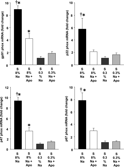
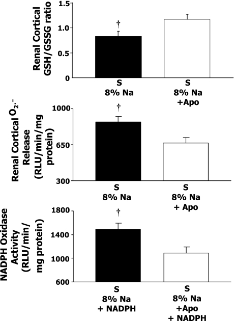
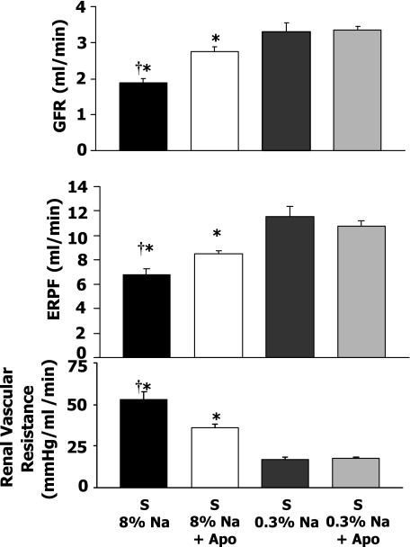
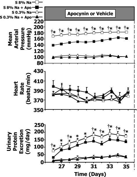
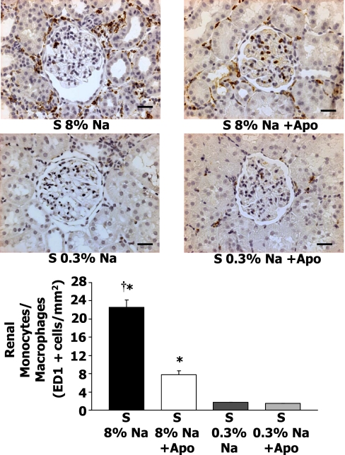
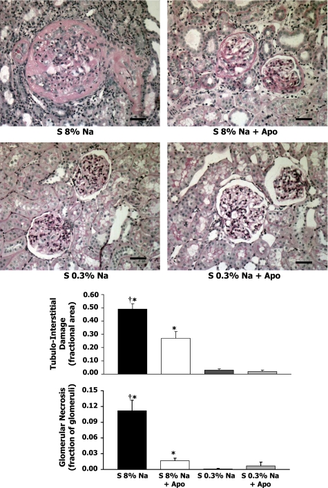
The goal of this study was to test the hypothesis that NADPH oxidase contributes importantly to renal cortical oxidative stress and inflammation, as well as renal damage and dysfunction, and increases in arterial pressure. Fifty-four 7- to 8-wk-old Dahl salt-sensitive (S) or R/Rapp strain rats were maintained for 5 wk on a high sodium (8%) or high sodium + apocynin (1.5 mmol/l in drinking water). Arterial and venous catheters were implanted on day 21. By day 35 in the high-Na S rats, mRNA expression of renal cortical gp91phox, p22phox, p47phox, and p67phox NADPH subunits in S rats increased markedly, and treatment of high-Na S rats with the NADPH oxidase inhibitor apocynin resulted in significant decreases in mRNA expression of these NADPH oxidase subunits. At the same time, in apocynin-treated S rats 1) renal cortical GSH/GSSG ratio increased, 2) renal cortical O2(.-) release and NADPH oxidase activity decreased, and 3) renal glomerular and interstitial damage markedly fell. Apocynin also decreased renal cortical monocyte/macrophage infiltration, and apocynin, but not the xanthine oxidase inhibitor allopurinol, attenuated decreases in renal hemodynamics and lowered arterial pressure. These data suggest that NADPH oxidase plays an important role in causing renal cortical oxidative stress and inflammation, which lead to decreases in renal hemodynamics, renal cortical damage, and increases in arterial pressure.


Figures

Fig. 1.
Fig. 1.
Effects of apocynin (Apo) and Na diet on the mRNA expression of NADPH oxidase subunits in Dahl salt-sensitive (S) rats in 8% Na (n = 9), 8% Na+Apo (n = 8), 0.3% Na (n = 6–7), and 0.3% Na+Apo groups (n = 6). †P < 0.05 when comparing 8% Na with 8% Na+Apo group. *P < 0.05 compared with 0.3% Na alone group.
Fig. 2.
Fig. 2.
Reduced-to-oxidized glutathione ratio (GSH/GSSG) in renal cortical tissue (8% Na, n = 9; 8% Na+Apo, n = 12), and renal cortical superoxide release and renal cortical NADPH oxidase activity in 8% Na (n = 13) and 8% Na+Apo (n = 9) in Dahl S rats. †P < 0.05 when comparing 8% Na with 8% Na+Apo group.
Fig. 3.
Fig. 3.
Glomerular filtration rate (GFR), renal plasma flow (ERPF), and renal vascular resistance responses in Dahl S rats in 8% Na (n = 9), 8% Na+Apo (n = 8), 0.3% Na (n = 7), and 0.3% Na+Apo (n = 7) groups. †P < 0.05 when comparing 8% Na with 8% Na+Apo group. *P < 0.05 compared with 0.3% Na alone group.
Fig. 4.
Fig. 4.
Mean arterial pressure, heart rate, and urinary protein excretion responses in Dahl S rats in 8% Na (n = 11), 8% Na+Apo (n = 9), 0.3% Na (n = 7), and 0.3% Na+Apo (n = 7) groups. †P < 0.05 when comparing 8% Na with 8% Na+Apo group. *P < 0.05 compared with 0.3% Na alone group.
Fig. 5.
Fig. 5.
Top: representative ED-1+cells (monocytes/macrophages) in the renal cortex of 8% Na or 0.3% Na Dahl S rats with and without Apo treatment. Kidneys were removed after 5 wk of the Na diet. Renal infiltration of monocytes/macrophages decreased in Dahl S rats treated with Apo. Bottom: average renal cortical monocyte/macrophage infiltration in Dahl S rats with 8% Na (n = 12), S 8% Na+Apo (n = 7), Dahl S rats with 0.3% Na (n = 7), and S 0.3% Na + Apo (n = 7) groups. †P < 0.05 when comparing 8% Na with 8% Na+Apo group. *P < 0.05 compared with 0.3% Na alone group. Scale bar = 30 μM.
Fig. 6.
Fig. 6.
Top: representative renal histology (all ×200 magnification) in the renal cortex of Dahl S rats with 8% Na (n = 12), S 8% Na+Apo (n = 7), Dahl S rats with 0.3% Na (n = 7), and S 0.3% Na+Apo (n = 7) groups. Bottom: average tubulointerstitial damage (fractional area) and glomerular necrosis (fraction of glomeruli). †P < 0.05 when comparing 8% Na with 8% Na+Apo group. *P < 0.05 compared with 0.3% Na alone group. Scale bar = 30 μM.

References

    1. Astern JM, Pendergraft WF III, Falk RJ, Jennette JC, Schmaier AH, Mahdi F, Preston GA. Myeloperoxidase interacts with endothelial cell-surface cytokeratin 1 and modulates bradykinin production by the plasma Kallikrein-Kinin system. Am J Pathol 171: 349–360, 2007. - PMC - PubMed
    1. Barton CH, Ni Z, Vaziri ND. Enhanced nitric oxide inactivation in aortic coarctation-induced hypertension. Kidney Int 60: 1083–1087, 2001. - PubMed
    1. Berber EY, Farber SJ, Earle DP Jr. Comparison of the constant infusion and urine collection techniques for the measurement of renal function. J Clin Invest 27: 710–719, 1948. - PubMed
    1. Beswick RA, Dorrance AM, Leite R, Webb RC. NADH/NADPH oxidase and enhanced superoxide production in the mineralocorticoid hypertensive rat. Hypertension 38: 1107–1111, 2001. - PubMed
    1. Beswick RA, Zhang H, Marable D, Catravas JD, Hill WD, Webb RC. Long-term antioxidant administration attenuates mineralocorticoid hypertension and renal inflammatory response. Hypertension 37: 781–786, 2001. - PubMed

Publication types

MeSH terms