. 2008 Oct;6(10):1521-33.
doi: 10.1158/1541-7786.MCR-07-2203.

Transforming growth factor-beta signaling-deficient fibroblasts enhance hepatocyte growth factor signaling in mammary carcinoma cells to promote scattering and invasion

Affiliations

Transforming growth factor-beta signaling-deficient fibroblasts enhance hepatocyte growth factor signaling in mammary carcinoma cells to promote scattering and invasion

Nikki Cheng et al. Mol Cancer Res. 2008 Oct.

Abstract

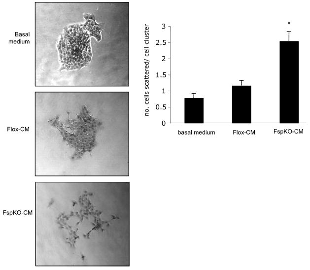
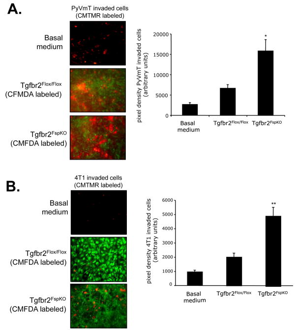
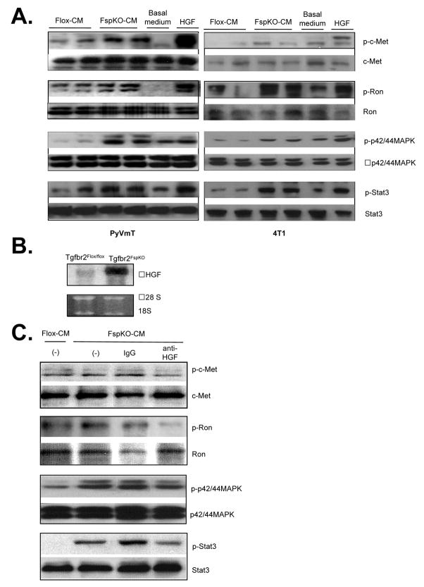
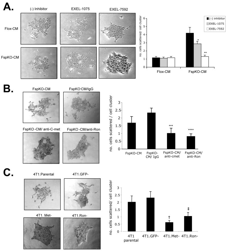
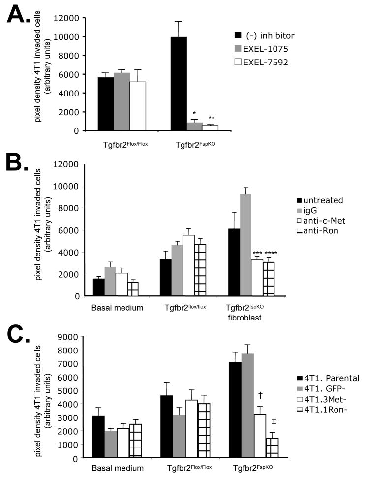
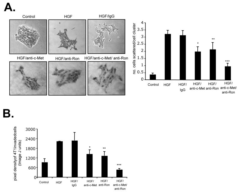
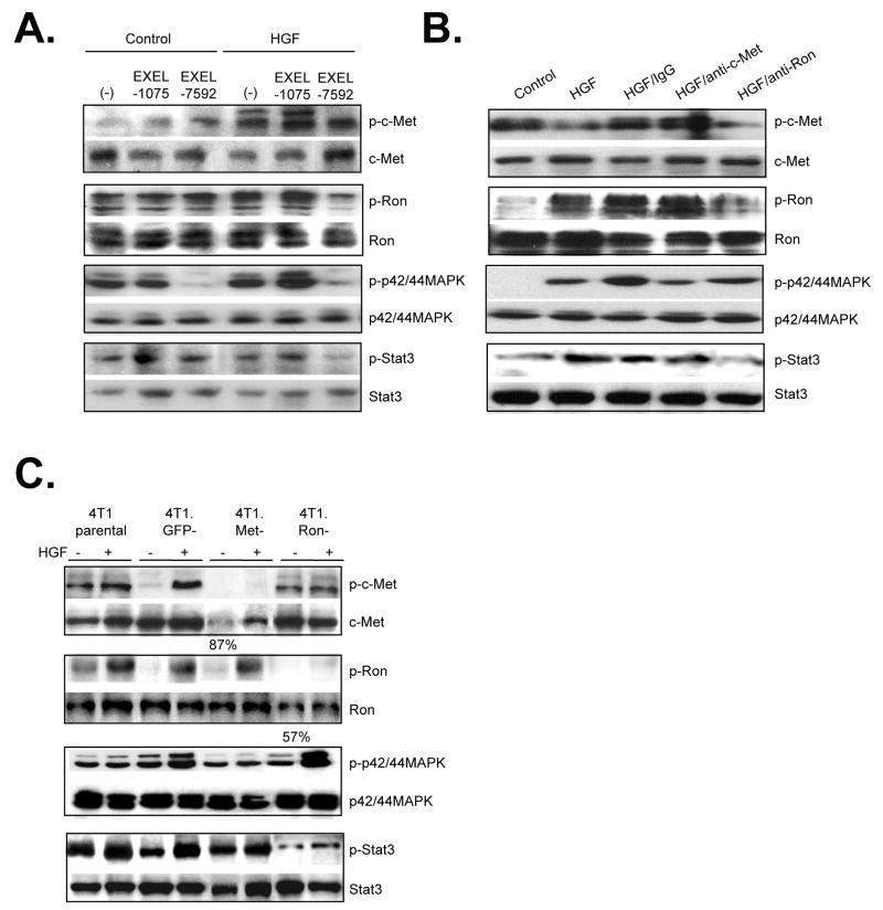
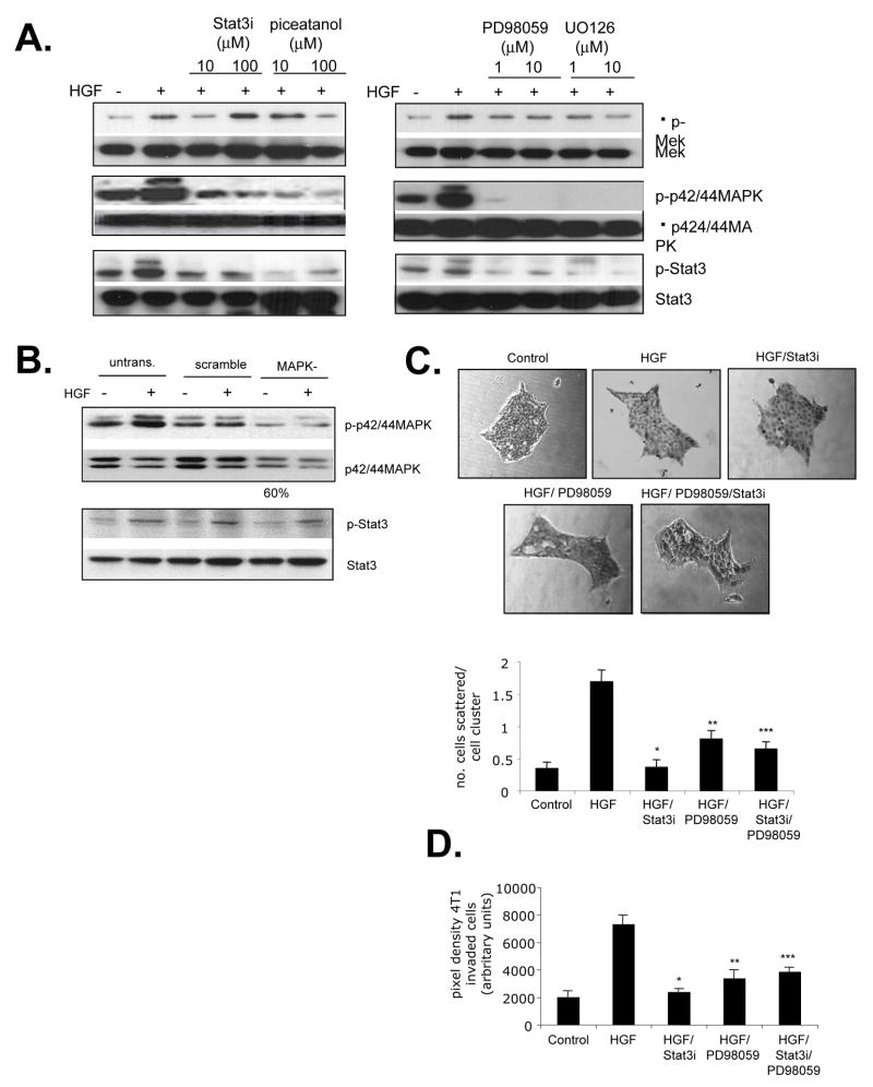
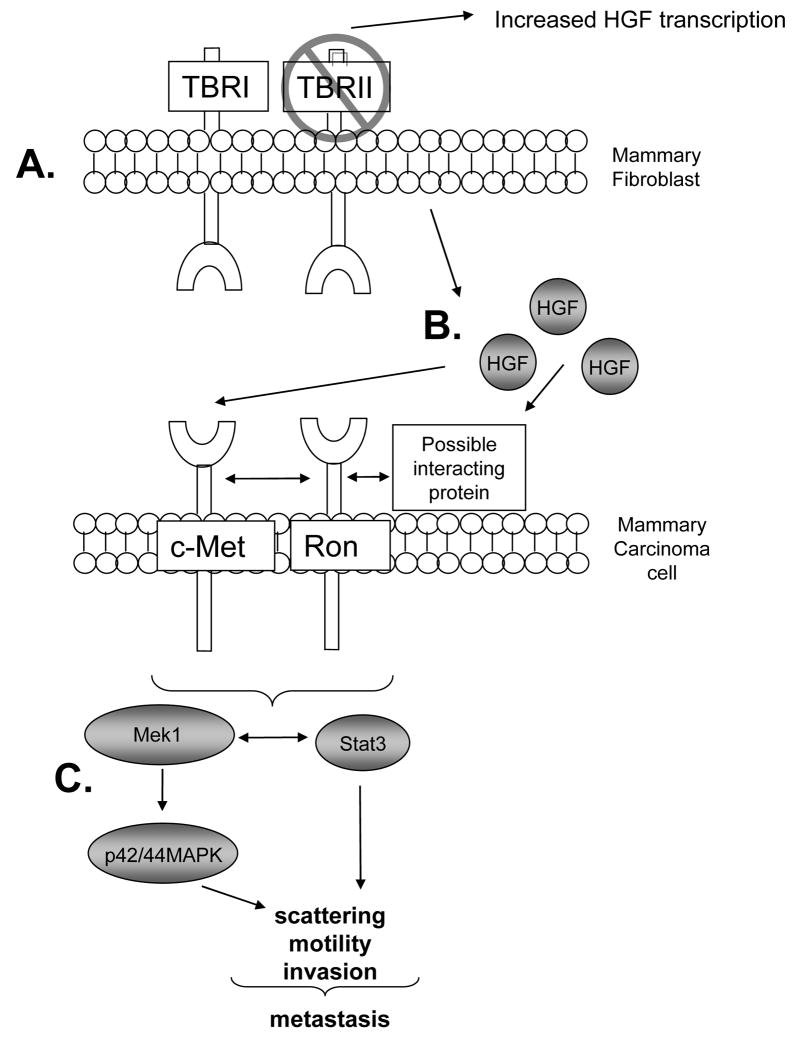
Fibroblasts are major cellular components of the tumor microenvironment, regulating tumor cell behavior in part through secretion of extracellular matrix proteins, growth factors, and angiogenic factors. In previous studies, conditional deletion of the type II transforming growth factor-beta (TGF-beta) receptor in fibroblasts (Tgfbr2FspKO) was shown to promote mammary tumor metastasis in fibroblast-epithelial cell cotransplantation studies in mice, correlating with increased expression of hepatocyte growth factor (HGF). Here, we advance our findings to show that Tgfbr2(FspKO) fibroblasts enhance HGF/c-Met and HGF/Ron signaling to promote scattering and invasion of mammary carcinoma cells. Blockade of c-Met and Ron by small interfering RNA silencing and pharmacologic inhibitors significantly reduced mammary carcinoma cell scattering and invasion caused by Tgfbr2FspKO fibroblasts. Moreover, neutralizing antibodies to c-Met and Ron significantly inhibited HGF-induced cell scattering and invasion, correlating with reduced Stat3 and p42/44MAPK phosphorylation. Investigation of the signal transducer and activator of transcription 3 (Stat3) and mitogen-activated protein kinase (MAPK) signaling pathways by pharmacologic inhibition and small interfering RNA silencing revealed a cooperative interaction between the two pathways to regulate HGF-induced invasion, scattering, and motility of mammary tumor cells. Furthermore, whereas c-Met was found to regulate both the Stat3 and MAPK signaling pathways, Ron was found to regulate Stat3 but not MAPK signaling in mammary carcinoma cells. These studies show a tumor-suppressive role for TGF-beta signaling in fibroblasts, in part by suppressing HGF signaling between mammary fibroblasts and epithelial cells. These studies characterize complex functional roles for HGF and TGF-beta signaling in mediating tumor-stromal interactions during mammary tumor cell scattering and invasion, with important implications in the metastatic process.


Conflict of interest statement

Conflict of interest: The authors have declared that no conflict of interest exists.

Figures

FIGURE 1
FIGURE 1. Tgfbr2FspKO fibroblasts promote mammary carcinoma cell scattering
4T1 cells were cultured in clusters of 50–75 cells and treated with basal medium (DMEM/F12/0.5% ABS) or conditioned medium fromTgfbr2Flox/Flox (Flox-CM) or Tgfbr2FspKO fibroblasts (FspKO-CM) for 24 hours. The number of cells scattered per cell cluster were counted. Statistical significance was determined by Two Tailed T-test; * vs. Flox-CM, p<0.05. Data are represented as mean±standard error of the mean (SEM).
FIGURE 2
FIGURE 2
Tgfbr2FspKO fibroblasts promote mammary carcinoma cell invasion. Fibroblasts were labeled with CMFDA (green) fluorochrome and plated on the underside of a Matrigel coated transwell membrane. A. PyVmT tumor cells or B. 4T1 cells were labeled with CMTMR (red) fluorochrome and plated on topside of the transwell filter in the presence or absence of Tgfbr2Flox/Flox control fibroblasts or Tgfbr2FspKO fibroblasts for 8 h. As a control, PyVmT and 4T1 tumor cells were seeded onto Matrigel coated transwell filters in basal medium containing DMEM/F12/0.5% ABS, in the absence of fibroblasts. Invaded tumor cells were visualized by fluorescence microscopy and micrographed at 20× magnification; an overlay of invaded carcinoma cells and fibroblasts is shown. Statistical significance was determined by Two Tailed T- test; *vs. Tgfbr2□lox/Flox fibroblasts, p<0.05, ** vs. Tgfbr2Flox/Flox fibroblasts, p<0.001. Data are represented as mean± SEM.
FIGURE 3
FIGURE 3
Conditioned medium from Tgfbr2FspKO fibroblasts enhances HGF signaling in mammary carcinoma cells. A. PyVmT (left panel) or 4T1 (right panel) carcinoma cells were treated with basal medium containing DMEM/F12/0.5% ABS, HGF (60 ng/ml) or fibroblast conditioned medium from Tgfbr2Flox/Flox control or Tgfbr2FspKO fibroblasts, and analyzed by western blot analysis for phosphorylation status of indicated proteins. Experiments were performed in quaduplicate with duplicate samples. Representative results are shown. B. Northern blot analysis of Tgfbr2Flox/Flox control and Tgfbr2FspKO fibroblasts. C. 4T1 cells were treated with fibroblast conditioned medium in the presence or absence of anti-HGF or control IgG and analyzed by western blot for the phosphorylation status of indicated proteins.
FIGURE 4
FIGURE 4
Inhibition of c-Met and Ron receptors block 4T1 cell scattering enhanced by Tgfbr2FspKO fibroblast conditioned medium. 4T1 cells were treated with basal medium (DMEM/F12/0.5% ABS), or conditioned medium from Tgfbr2Flox/flox (Flox-CM) or Tgfbr2FspKO fibroblasts (FspKO-CM) in the presence or absence of the following c-Met or Ron inhibitors for 24 hours: A. Treatment of cells with 250 nM EXEL-7592 (VEGFR/c-Met inhibitor) or EXEL-1075 (VEGFR control inhibitor). B. 4T1 cells were treated with 1 μg/ml anti-c-Met, anti-Ron or control IgG. C. siRNA silencing of c-Met, Ron or control GFP, treatment with FspKO-CM. Quantitation of the expression of c-Met and Ron by densitometry analysis of western blots indicated an 87% reduction in c-Met and 57% reduction in Ron expression relative to expression in 4T1 Parental cells. Statistical significance was determined by two tailed T-test: * vs FspKO-CM; p=0.12, ** vs. FspKO-CM/EXEL1075; p<0.01, *** vs. FspKO-CM/IgG; p<0.01, **** vs FspKO-CM/IgG; p<0.01, vs. 4T1.GFP-; p<0.01, vs. 4T1.GFP-; p<0.01. Data are represented as mean± SEM.
FIGURE 5
FIGURE 5
Inhibition of c-Met and Ron receptors block 4T1 carcinoma cell invasion induced by Tgfbr2Fspko fibroblasts. 4T1 cells labeled with CMTMR CellTracker dye were seeded onto Matrigel coated transwell membrane coated with Tgfbr2Flox/Flox or Tgfbr2FspKO fibroblasts labeled with CMFDA CellTracker dye and assessed for invasion after 8 h in the presence or absence of the following c-Met and Ron inhibitors: A. 250 nM EXEL-7592 or EXEL-1075. B. 1 μg/ml anti-c-Met, anti-Ron or control goat IgG. C. siRNA silencing of c-Met, Ron or control GFP. Experiments were performed in replicates of four and repeated in triplicate. Quantitation of carcinoma cells was determined by measuring pixel density of CMTMR labeled cells using Image J software (arbitrary units). Statistical significance was determined by Two Tailed T-test: * vs. Tgfbr2FspKO fibroblasts/(−) inhibitor, p<0.001; **vs. Tgfbr2FspKO fibroblasts/(−) inhibitor, p<0.001; ***vs Tgfbr2FspKO fibroblasts/IgG, p<0.01; ****vs Tgfbr2FspKO fibroblasts/IgG, p<0.01; vs. 4T1.GFP-/Tgfbr2FspKO fibroblasts, p<0.05; vs. 4T1.GFP-/Tgfbr2FspKO fibroblasts, p<0.05. Data are represented as mean±SEM.
FIGURE 6
FIGURE 6
Ron and c-Met receptors cooperate to mediate HGF- induced mammary tumor cell scattering, motility and invasion. 4T1 tumor cells were treated with Control (serum free medium) or HGF in the presence or absence of control goat IgG (1μg/ml) or neutralizing antibodies (1μg/ml) to c- Met and Ron, and assessed for the ability to A. scatter B. invade across a Matrigel coated membrane. Pixel density of invaded cells was measured by Image J software. Statistical significance was determined by Two Tailed T-test: *vs. HGF/IgG; p<0.01, **vs. HGF/IgG; p<0.01, and Two Way ANOVA Test: ***vs. HGF/anti-c-Met and HGF/anti-Ron treatments; p<0.05. Data are represented as mean±SEM.
FIGURE 7
FIGURE 7
Ron and c-Met receptors signal independently to activate MAPK and Stat3 pathways in 4T1 carcinoma cells. 4T1 cells were treated with control (serum free medium) or HGF in the presence of absence of the following c-Met and Ron inhibitors and analyzed by western blot analysis for the phosphorylated status of indicated proteins: A. Treatment of cells with 250 nM EXEL-7592 or control KDR inhibitor EXEL-1075 B. Treatment of cells with neutralizing antibodies to c-Met and Ron. C. siRNA silencing of c-Met and Ron. Expression of c-Met and Ron was quantified by densitometry analysis of western blots and is shown as a percentage of expression relative to c-Met and Ron expression in 4T1 Parental cells. Representative results of triplicate experiments are shown.
FIGURE 8
FIGURE 8
Stat3 and p42/44 MAPK pathways cooperate to mediate HGF induced invasion and scattering A. 4T1 tumor cells were treated with indicated doses of the Stat3 inhibitors: Stat3 inhibitor peptide or piceatanol and indicated doses of the MAPK inhibitors PD98059 or U0126 and analyzed by western blot for the phosphorylation status of the indicated proteins. B. p42/44MAPK expression was inhibited in 4T1 cells by siRNA silencing. 4T1 cells were treated with HGF and analyzed by western blot analysis for the phosprhoylatin status of indicated proteins. C and D: 4T1 cells were treated with Control (serum free medium), HGF (60 ng/ml) in the presence or absence of 10 μM Stat3 peptide inhibitor (Stat3i) or 1 μM PD98059, and analyzed for the ability to C. scatter and D. invade through a Matrigel coated membrane. Statistical significance was determined by Two T-tailed T-test: * vs. HGF; p<0.01, ** vs. HGF; p<0.01 and Two Way ANOVA: *** vs. HGF/PD98059 and HGF/Stat3i treated cells; p>0.05. Data are represented as mean±SEM.
FIGURE 9
FIGURE 9
Model of TGF-β and HGF signaling in fibroblast: tumor interactions. A. Cre mediated deletion of the TGF-β type II receptor in mammary fibroblasts inhibits TGF-β signaling, resulting in upregulated HGF expression at the RNA level as well as protein secretion [17,26] B. HGF binds to c-Met directly as may interact with Ron indirectly to enhance c-Met and Ron mediated downstream signaling in mammary epithelial cells. C. Increased cooperative interactions between Stat3 and p42/44MAPK signaling results in mammary carcinoma cell scattering, motility and invasion, important processes involved in metastatic spread.

References

    1. Bhowmick NA, Moses HL. Tumor-stroma interactions. Curr Opin Genet Dev. 2005;15:97–101. - PMC - PubMed
    1. Liotta LA, Kohn EC. The microenvironment of the tumour-host interface. Nature. 2001;411:375–379. - PubMed
    1. Kurose K, Gilley K, Matsumoto S, Watson PH, Zhou XP, Eng C. Frequent somatic mutations in PTEN and TP53 are mutually exclusive in the stroma of breast carcinomas. Nat Genet. 2002;32:355–357. - PubMed
    1. Barcellos-Hoff MH, Ravani SA. Irradiated mammary gland stroma promotes the expression of tumorigenic potential by unirradiated epithelial cells. Cancer Res. 2000;60:1254–1260. - PubMed
    1. Camps J, Chang S, Hsu T, Freeman M, hong S, Zhau H, von Eschenbach A, Chung L. Fibroblast-mediated acceleration of human epithelial tumor growth in vivo. Proc Natl Acad Sci U S A. 1990;87:75–79. - PMC - PubMed

Publication types

MeSH terms

Substances