. 2009 Jan 15;18(2):227-40.
doi: 10.1093/hmg/ddn339. Epub 2008 Oct 15.

FGFR3 promotes synchondrosis closure and fusion of ossification centers through the MAPK pathway

Affiliations

FGFR3 promotes synchondrosis closure and fusion of ossification centers through the MAPK pathway

Takehiko Matsushita et al. Hum Mol Genet. .

Abstract

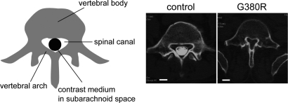
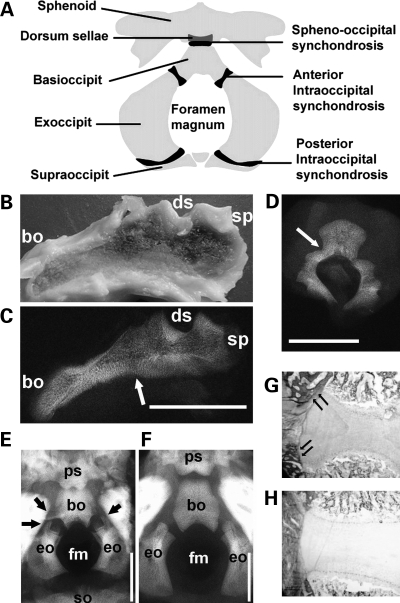
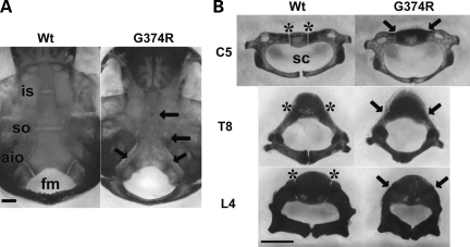
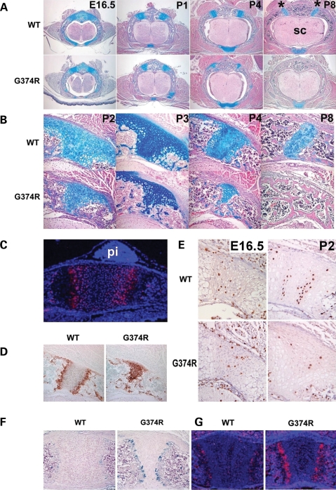
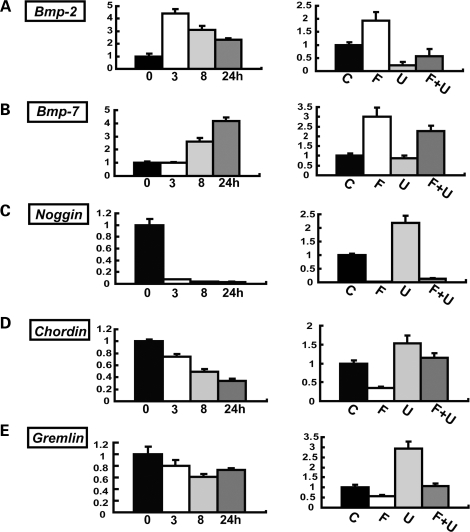
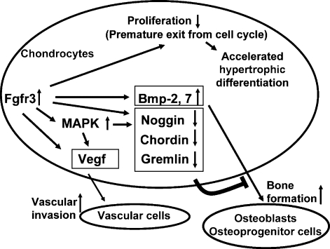
Activating mutations in FGFR3 cause achondroplasia and thanatophoric dysplasia, the most common human skeletal dysplasias. In these disorders, spinal canal and foramen magnum stenosis can cause serious neurologic complications. Here, we provide evidence that FGFR3 and MAPK signaling in chondrocytes promote synchondrosis closure and fusion of ossification centers. We observed premature synchondrosis closure in the spine and cranial base in human cases of homozygous achondroplasia and thanatophoric dysplasia as well as in mouse models of achondroplasia. In both species, premature synchondrosis closure was associated with increased bone formation. Chondrocyte-specific activation of Fgfr3 in mice induced premature synchondrosis closure and enhanced osteoblast differentiation around synchondroses. FGF signaling in chondrocytes increases Bmp ligand mRNA expression and decreases Bmp antagonist mRNA expression in a MAPK-dependent manner, suggesting a role for Bmp signaling in the increased bone formation. The enhanced bone formation would accelerate the fusion of ossification centers and limit the endochondral bone growth. Spinal canal and foramen magnum stenosis in heterozygous achondroplasia patients, therefore, may occur through premature synchondrosis closure. If this is the case, then any growth-promoting treatment for these complications of achondroplasia must precede the timing of the synchondrosis closure.


Figures

Figure 1.
Figure 1.
Spinal canal stenosis in achondroplasia. Axial CT myelogram of the fifth lumbar spine shows the narrowing of the spinal canal in a 46-year-old female achondroplasia patient with FGFR3 G380R mutation, who presented with paraparesis (right). The contrast medium injected into the subarachnoid space was excluded in the achondroplasia patient due to severe spinal canal stenosis. (Left) 41-year-old female patient with irrelevant spinal disorder. White bars indicate 1 cm.
Figure 2.
Figure 2.
The spine and cranial base of homozygous achondroplasia and thanatophoric dysplasia. (A) An illustration of the anatomy of the cranial base. (BD) Cranial base and lumbar spine of homozygous achondroplasia (Case 1). Gross appearance (B) and X-ray (C) of mid-sagittal slice of the cranial base of homozygous achondroplasia. Arrow indicates the fused spheno-occipital synchondrosis. (D) The third lumbar spine showed complete fusion of the neurocentral synchondrosis on one side and partial fusion on the other side. (E and F) Cranial view of the cranial base of a thanatophoric dysplasia fetus (E) (Case 2) at 27 weeks of gestation and a control fetus (F) (Case 3) at 26 weeks of gestation without any signs of skeletal dysplasia. The anterior intraoccipital synchondroses of the thanatophoric dysplasia fetus are being encased in bone (arrows). (G and H) Goldner staining of the spheno-occipital synchondrosis of Case 2 (G) and Case 3 (H), showing increased bone formation in thanatophoric dysplasia (arrows). Scale bars indicate 10 mm. Abbreviations: bo, basioccipital; eo, exoccipital; fm, foramen magnum; so, supraoccipital; sp, sphenoid; ds, dorsum sellae; ps, postsphenoid.
Figure 3.
Figure 3.
Skeletal preparations after alizarin red staining of mice that express Fgfr3 G374R and wild-type littermate mice at P10. Synchondroses in the cranial base (A) and spine (B) were prematurely closed in mice that express Fgfr3 G374R. Arrows indicate fused synchondroses. Asterisks indicate the neurocentral synchondroses. Scale bars indicate 1 mm. Abbreviations: Wt, wild-type mice, (G374R) mice that express Fgfr3 G374R; is, intersphenoid; so, spheno-occipital; aio, anterior intraoccipital; sc, spinal canal; C5, 5th cervical vertebrae; T8, eighth thoracic vertebrae; L4, fourth lumbar vertebrae.
Figure 4.
Figure 4.
Premature loss of proliferating chondrocytes and increased vascular invasion in the synchondroses of mice that express Fgfr3 G374R. (A) Hematoxylin, eosin and alcian blue-stained horizontal sections of the spine showed premature closure of the neurocentral synchondroses in mice that express Fgfr3 G374R. Asterisk indicates neurocentral synchondroses and sc denotes spinal cord. (B) Higher magnification of the closing neurocentral synchondroses between P2 and P8. All the vertebrae shown in (A) and (B) are either T12 or L1. (C) In situ hybridization of Fgfr3 of the spheno-occipital synchondrosis of wild-type mice at P1. Fgfr3 is strongly expressed in chondrocytes in the proliferating and prehypertrophic zones. pi denotes pituitary glands. (D) Type X collagen immunostaining of neurocentral synchondrosis of mice that express Fgfr3 G374R and wild-type littermate mice at P4. Mice that express Fgfr3 G374R showed premature loss of the zones of resting and proliferating chondrocytes. (E) Immunostaining of the neurocentral synchondrosis for BrdU at E16.5 and P2. BrdU-labeled chondrocytes are markedly reduced in the closing neurocentral synchondrosis of mice that express Fgfr3 G374R at P2. (F) Immunostaining for von Willebrand factor showed increased vascular endothelial cells at the chondro-osseous junction of the sternum of mice that express Fgfr3 G374R at P5. (G) In situ hybridization analysis showed increased Vegf expression in the synchondroses of the sternum of mice that express Fgfr3 G374R at P1.
Figure 5.
Figure 5.
Increased osteoblast differentiation and bone formation in mice that express Fgfr3 G374R. (A) Increased number of X-gal-stained osteoblasts around the neurocentral synchondroses of the lower thoracic spine in Fgfr3 G374R mice at P3. Zp3-Cre transgene was used to express Fgfr3 G374R in a systemic manner. Col1a1-LacZ transgene was used to express LacZ in osteoblasts. (B) Immunohistochemistry for Runx2 showed an increase in Runx2-positive cells in the periosteum flanking the neurocentral synchondroses of Fgfr3 G374R mice at P4. Right panels show magnified images of the boxed areas in the left panels. (C and D) X-gal staining of the neurocentral synchondroses of the lower thoracic spine (C) and sternum (D) at P3. Col2a1-Cre transgene was used to express Fgfr3 G374R in chondrocytes of Col1a1-LacZ transgenic mice. (E) Chondrocyte and osteoblast-specific recombination was confirmed by crossing Col2a1-Cre and Col1a1-Cre transgenic mice with the ROSA26 reporter mice. X-gal staining of the sternum showed chondrocyte-specific recombination by the Col2a1-Cre transgene at P2 and osteoblast-specific recombination by the Col1a1-Cre transgene at P3. (F and G) Hematoxylin, eosin and alcian blue-stained horizontal sections of the lower thoracic spine at P11 (F) and P3 (G). (F) No acceleration of synchondrosis closure in Fgfr3 G374R mice, in which the Col1a1-Cre transgene was used to recombine Fgfr3 in osteoblasts. (G) Premature synchondrosis closure (arrows) in Fgfr3 G374R mice, in which the Col2a1-Cre transgene was used to recombine Fgfr3 in chondrocytes.
Figure 6.
Figure 6.
Premature synchondrosis closure in mice that express a constitutively active mutant of MEK1 (S218/222E, Δ32-51) in chondrocytes. Skeletal preparations of the (A) cranial base and (B) spine showed premature synchondrosis closure. Arrows indicate fused synchondroses. Abbreviations: is, intersphenoid; so, spheno-occipital; aio, anterior intraoccipital; asterisk, neurocentral synchondroses; C5, fifth cervical vertebrae; T8, eighth thoracic vertebrae. (C) Upper panels show hematoxylin, eosin and alcian blue (HE Al B) staining of the spheno-occipital synchondrosis at P1. In transgenic mice that express a constitutively active mutant of MEK1 in chondrocytes, the eosin-stained bone cortex (green arrow) over the synchondrosis connects the cortices of the sphenoid and occipital bones, whereas in wild type mice, the synchondrosis is flanked by the thin perichondrium. Lower panels show in situ hybridization for Bone sialoprotein (BSP) of neighboring sections. While the perichondrium flanking the synchondrosis did not express Bone sialoprotein in wild type mice, mice that express a constitutively active mutant of MEK1 in chondrocytes showed intense signal for Bone sialoprotein over the synchondrosis (green arrow), indicating accelerated osteoblast differentiation in the perichondrium.
Figure 7.
Figure 7.
FGF18 upregulates Bmp-2, Bmp-7 and downregulates Noggin, Chordin and Gremlin in primary chondrocytes. (A) Bmp-2, (B) Bmp-7, (C) Noggin, (D) Chordin and (E) Gremlin. For each gene, the left panel shows the time course of gene expression levels after stimulation with 20 ng/ml FGF18. Total RNA was extracted at 3, 8 and 24 h after stimulation. mRNA expression levels were examined by real-time PCR. The right panel shows the effects of U0126 on gene expression. Primary chondrocytes were treated with 20 ng/ml FGF18 in the presence or absence of 20 µm U0126. (C) Control, (F) FGF18, (U) U0126, (F + U) FGF18 and U0126. Total RNA was extracted at 3 h after FGF18 stimulation for Bmp-2 and at 24 h for Bmp-7, Noggin, Chordin and Gremlin. In all panels, mRNA levels were normalized by the values for the control culture harvested at each time point. Data represent mean ± SD. The figure presents data from one of two experiments that produced similar results.
Figure 8.
Figure 8.
Decreased Noggin and Gremlin expression in chondrocytes of mice that express Fgfr3 G374R. (A) In situ hybridization analysis showed reduced Noggin expression in the neurocentral synchondroses in the lower thoracic spine of mice that express Fgfr3 G374R at P1. The boxed area is magnified on the left. Neighboring sections were stained with alcian blue. (B) In situ hybridization analysis showed decreased Noggin expression in the spheno-occipital synchondrosis of mice that express a constitutively active mutant MEK1(S218/222E, Δ32-51) in chondrocytes compared with wild-type littermate mice at P1. Alcian blue staining of the neighboring sections are presented in Fig. 6C. (C) In situ hybridization analysis showed reduced Gremlin expression in the synchondroses in the sternum of mice that express Fgfr3 G374R at P1. Neighboring sections were stained with alcian blue staining. (DF) Real-time PCR analysis showed reduced Gremlin (D) and Noggin (E) expression and increased Bmp-7 (F) expression in the epiphyseal cartilage of mice that express Fgfr3 G374R (n = 3) compared with wild-type littermate mice (n = 3) at P3. Total RNA was extracted from the epiphyseal cartilage of long bones. The expression of each gene was normalized by the expression level in wild type mice. Data represent mean ± SD. The figure presents representative data from repeated experiments. *P < 0.05, **P < 0.02 (unpaired Student's t-test).
Figure 9.
Figure 9.
Model whereby increased Fgfr3 signaling in chondrocytes causes premature synchondrosis closure and unification of ossification centers. Increased Fgfr3 signaling induces premature exit from the cell cycle and accelerates the transition into hypertrophic chondrocytes. Increased Fgfr3 signaling also causes upregulation of Vegf in chondrocytes, promoting vascular invasion. Increased secretion of Bmp ligands and decreased secretion of Bmp antagonists result in enhanced osteoblast differentiation of osteoprogenitors in the periosteum, promoting bone formation and fusion of ossification centers. The upregulation of Vegf and Bmps and downregulation of Bmp antagonists are at least partially mediated by the MAPK pathway.

Similar articles

Cited by

References

    1. van der Eerden B.C., Karperien M., Wit J.M. Systemic and local regulation of the growth plate. Endocr. Rev. 2003;24:782–801. - PubMed
    1. Nilsson O., Marino R., De Luca F., Phillip M., Baron J. Endocrine regulation of the growth plate. Horm. Res. 2005;64:157–165. - PubMed
    1. Peters K., Ornitz D., Werner S., Williams L. Unique expression pattern of the FGF receptor 3 gene during mouse organogenesis. Dev. Biol. 1993;155:423–430. - PubMed
    1. Delezoide A.L., Benoist-Lasselin C., Legeai-Mallet L., Le Merrer M., Munnich A., Vekemans M., Bonaventure J. Spatio-temporal expression of FGFR 1, 2 and 3 genes during human embryo-fetal ossification. Mech. Dev. 1998;77:19–30. - PubMed
    1. Rousseau F., Bonaventure J., Legeai-Mallet L., Pelet A., Rozet J.M., Maroteaux P., Le Merrer M., Munnich A. Mutations in the gene encoding fibroblast growth factor receptor-3 in achondroplasia. Nature. 1994;371:252–254. - PubMed

Publication types

MeSH terms

Substances