Comparative Study
. 2008 Oct 15;28(42):10460-71.
doi: 10.1523/JNEUROSCI.2518-08.2008.

Distinct roles of PDE4 and PDE10A in the regulation of cAMP/PKA signaling in the striatum

Affiliations
Comparative Study

Distinct roles of PDE4 and PDE10A in the regulation of cAMP/PKA signaling in the striatum

Akinori Nishi et al. J Neurosci. .

Abstract

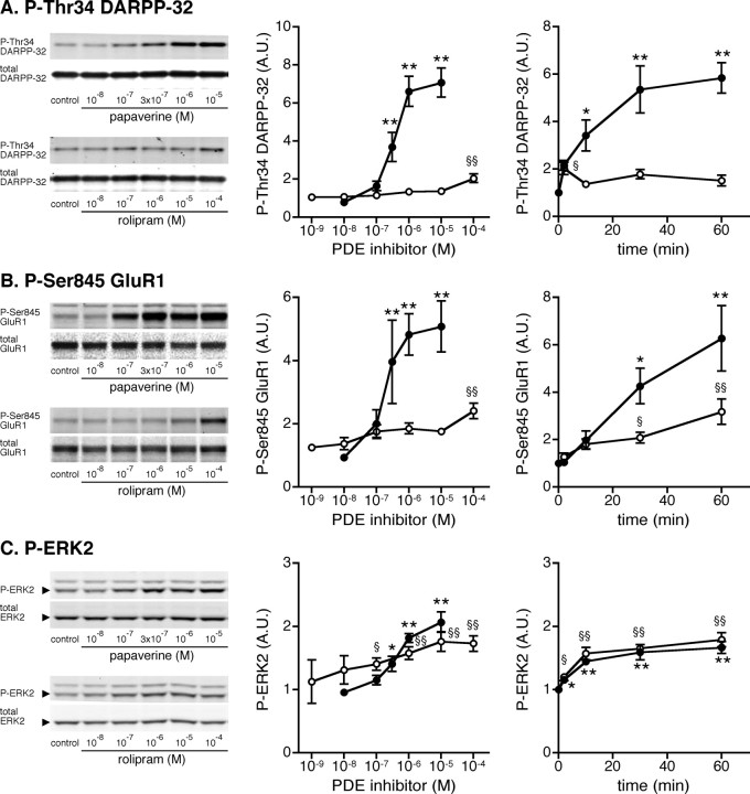
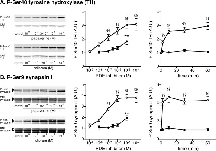
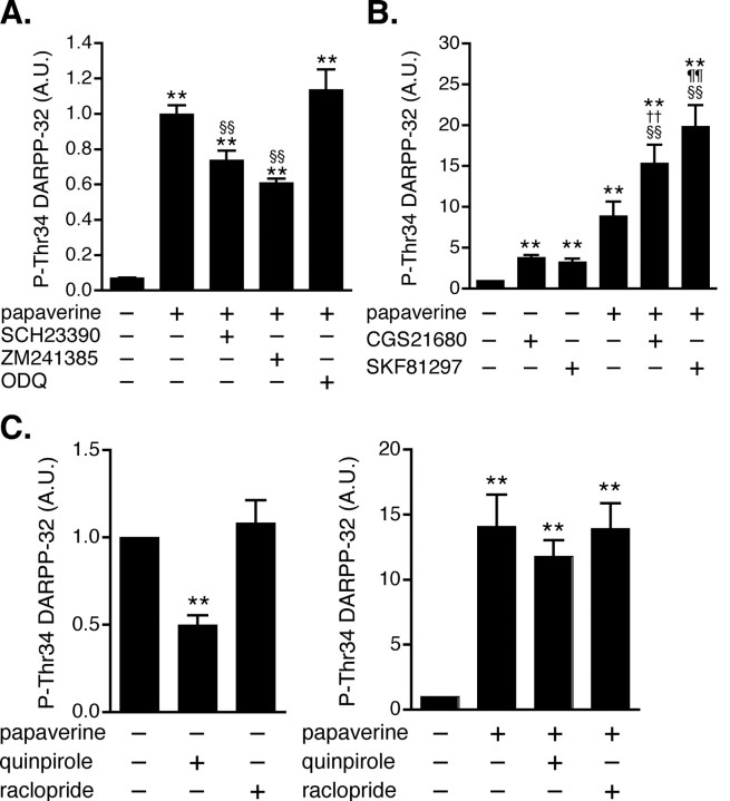
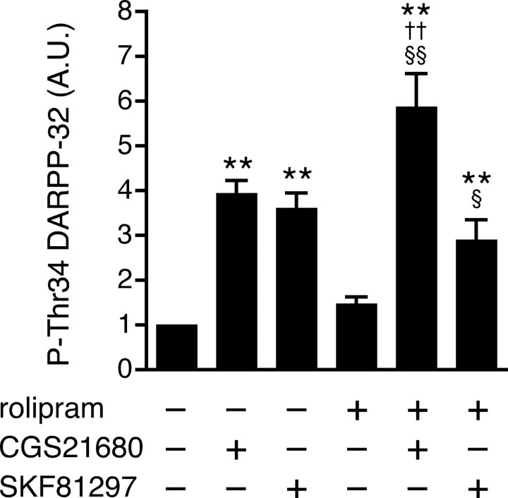
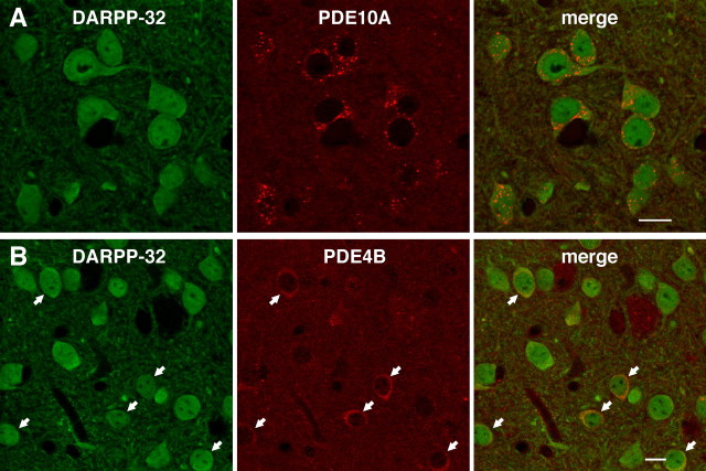
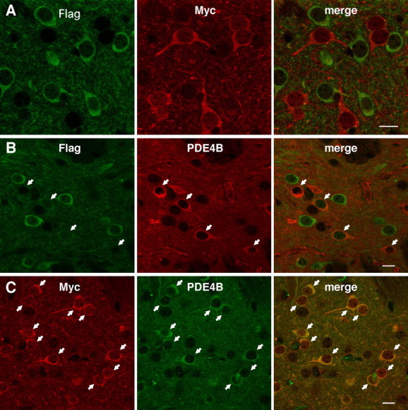
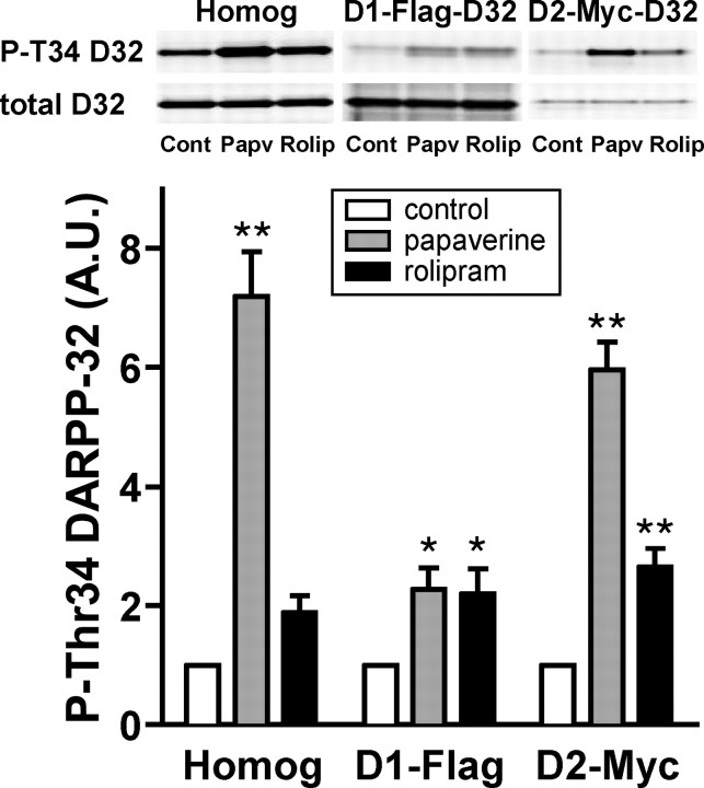
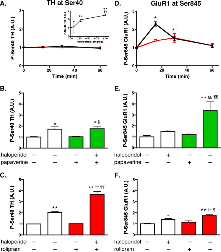
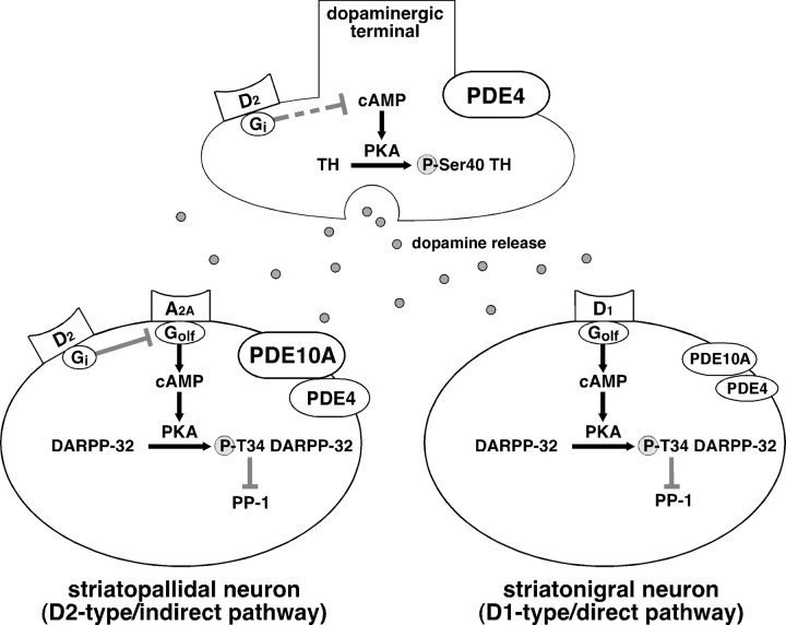
Phosphodiesterase (PDE) is a critical regulator of cAMP/protein kinase A (PKA) signaling in cells. Multiple PDEs with different substrate specificities and subcellular localization are expressed in neurons. Dopamine plays a central role in the regulation of motor and cognitive functions. The effect of dopamine is largely mediated through the cAMP/PKA signaling cascade, and therefore controlled by PDE activity. We used in vitro and in vivo biochemical techniques to dissect the roles of PDE4 and PDE10A in dopaminergic neurotransmission in mouse striatum by monitoring the ability of selective PDE inhibitors to regulate phosphorylation of presynaptic [e.g., tyrosine hydroxylase (TH)] and postsynaptic [e.g., dopamine- and cAMP-regulated phosphoprotein of M(r) 32 kDa (DARPP-32)] PKA substrates. The PDE4 inhibitor, rolipram, induced a large increase in TH Ser40 phosphorylation at dopaminergic terminals that was associated with a commensurate increase in dopamine synthesis and turnover in striatum in vivo. Rolipram induced a small increase in DARPP-32 Thr34 phosphorylation preferentially in striatopallidal neurons by activating adenosine A(2A) receptor signaling in striatum. In contrast, the PDE10A inhibitor, papaverine, had no effect on TH phosphorylation or dopamine turnover, but instead robustly increased DARPP-32 Thr34 and GluR1 Ser845 phosphorylation in striatal neurons. Inhibition of PDE10A by papaverine activated cAMP/PKA signaling in both striatonigral and striatopallidal neurons, resulting in potentiation of dopamine D(1) receptor signaling and inhibition of dopamine D(2) receptor signaling. These biochemical results are supported by immunohistochemical data demonstrating differential localization of PDE10A and PDE4 in striatum. These data underscore the importance of individual brain-enriched cyclic-nucleotide PDE isoforms as therapeutic targets for neuropsychiatric and neurodegenerative disorders affecting dopamine neurotransmission.


Figures

Figure 1.
Figure 1.
Effect of a PDE10A inhibitor, papaverine, and a PDE4 inhibitor, rolipram, on DARPP-32, GluR1, and ERK2 phosphorylation in neostriatal slices. A–C, Mouse neostriatal slices were treated with various concentrations of papaverine (closed circles) or rolipram (open circles) for 60 min (left and center panels) and with papaverine (1 μm; closed circles) or rolipram (10 μm; open circles) for the indicated times (right panels). Changes in the phosphorylation of DARPP-32 at Thr34 (the PKA site), GluR1 at Ser845 (the PKA site), and ERK at Thr202/Tyr204 (the MEK site) were determined by Western blotting using phosphorylation-state-specific antibodies. Typical immunoblots are shown in left panels. Data represent means ± SEM for 5–13 experiments. *p < 0.05, **p < 0.01 compared with untreated slices for papaverine; § p < 0.05, §§ p < 0.01 compared with untreated slices for rolipram; one-way ANOVA followed by Newman–Keuls test.
Figure 2.
Figure 2.
Effect of papaverine and rolipram on TH and synapsin I phosphorylation in neostriatal slices. A–C, Mouse neostriatal slices were treated with various concentrations of papaverine (closed circles) or rolipram (open circles) for 60 min (left and center panels) and with papaverine (1 μm; closed circles) or rolipram (10 μm; open circles) for the indicated times (right panels). Changes in the phosphorylation of TH at Ser40 (the PKA site) and synapsin I at Ser9 (the PKA/CaMKI site), which are selectively expressed at presynaptic terminals, were determined by Western blotting using phosphorylation-state-specific antibodies. Typical immunoblots are shown in left panels. Data represent means ± SEM for 5–13 experiments. **p < 0.01 compared with untreated slices for papaverine; §§ p < 0.01 compared with untreated slices for rolipram; one-way ANOVA followed by Newman–Keuls test.
Figure 3.
Figure 3.
Effect of papaverine on dopamine D1, dopamine D2, and adenosine A2A receptor signaling in neostriatal slices. A, The effect of papaverine (10 μm for 60 min) on DARPP-32 Thr34 phosphorylation was examined in the presence of a dopamine D1 receptor antagonist, SCH23390 (1 μm for 60 min), an adenosine A2A receptor antagonist, ZM241385 (1 μm for 60 min), or an inhibitor of soluble guanylyl cyclase, ODQ (10 μm for 60 min). Data represent means ± SEM for 4–6 experiments. B, In slices pretreated with papaverine (10 μm for 60 min) and adenosine deaminase (30 μg/ml for 60 min), the effects of a dopamine D1 agonist, SKF81297 (1 μm for 5 min) and an adenosine A2A receptor agonist, CGS21680 (5 μm for 2 min), on DARPP-32 Thr34 phosphorylation were examined. Adenosine deaminase, additionally included in the incubation medium to decease tissue content of adenosine, reduced the basal and papaverine-induced levels of phospho-Thr34 DARPP-32. Data represent means ± SEM for 6–19 experiments. C, The effect of a dopamine D2 receptor agonist, quinpirole (1 μm for 10 min), and a dopamine D2 receptor antagonist, raclopride (1 μm for 10 min), on DARPP-32 Thr34 phosphorylation was examined in the absence (left) or presence (right) of papaverine (10 μm for 70 min). Data represent means ± SEM for 7–13 experiments. **p < 0.01 compared with control; §§ p < 0.01 compared with papaverine alone; †† p < 0.01 compared with CGS21680 alone; ¶¶ p < 0.01 compared with SKF81297 alone; one-way ANOVA followed by Newman–Keuls test.
Figure 4.
Figure 4.
Effect of rolipram on dopamine D1 and adenosine A2A receptor signaling in neostriatal slices. In slices pretreated with rolipram (100 μm for 60 min), the effects of a dopamine D1 agonist, SKF81297 (1 μm for 5 min), and an adenosine A2A receptor agonist, CGS21680 (5 μm for 2 min), on DARPP-32 Thr34 phosphorylation were examined. Data represent means ± SEM for 6–19 experiments. **p < 0.01 compared with control; § p < 0.05, §§ p < 0.01 compared with rolipram alone; †† p < 0.01 compared with CGS21680 alone; one-way ANOVA followed by Newman–Keuls test.
Figure 5.
Figure 5.
Expression of PDE4B and PDE10A in the striatum. A, B, Double immunostaining of striatal tissues with (A) DARPP-32 and PDE10A antibodies and (B) DARPP-32 and PDE4B antibodies. Arrows in B indicate DARPP-32-positive neurons with strong PDE4B immunoreactivity. Scale bars, 10 μm.
Figure 6.
Figure 6.
High expression of PDE4B in Myc-positive, striatopallidal neurons in the striatum of D1-DARPP-32-Flag/D2-DARPP-32-Myc mutant mice. A, Expression of Flag- and Myc-tagged DARPP-32 in striatonigral and striatopallidal neurons, respectively, in the striatum of D1-DARPP-32-Flag/D2-DARPP-32-Myc mutant mice. B, Double immunostaining of striatal tissues from D1-DARPP-32-Flag/D2-DARPP-32-Myc mice with Flag and PDE4B antibodies. C, Double immunostaining of striatal tissues from D1-DARPP-32-Flag/D2-DARPP-32-Myc mice with Myc and PDE4B antibodies. Striatal neurons with strong PDE4B immunoreactivity, indicted by arrows, correspond to Myc-positive, striatopallidal neurons. Scale bars, 10 μm.
Figure 7.
Figure 7.
Neuronal type-specific regulation of DARPP-32 phosphorylation by papaverine and rolipram in neostriatal slices from D1-DARPP-32-Flag/D2-DARPP-32-Myc mice. Neostriatal slices from D1-DARPP-32-Flag/D2-DARPP-32-Myc mice were incubated with papaverine (10 μm) or rolipram (100 μm) for 60 min. Flag-tagged DARPP-32, expressed in D1 receptor-enriched striatonigral neurons, and Myc-tagged DARPP-32, expressed in D2 receptor-enriched striatopallidal neurons, were immunoprecipitated. The figure shows data from total striatal homogenate (Homog), Flag-tagged DARPP-32 in striatonigral neurons (D1-Flag), and Myc-tagged DARPP-32 in striatopallidal neurons (D2-Myc). Typical immunoblots for detection of phospho-Thr34 DARPP-32 (P-T34 D32) and total DARPP-32 (total D32) in the same membrane are shown at the top. The levels of phospho-Thr34 DARPP-32 and total DARPP-32 were quantified by the Odyssey infrared imaging system, and the data (phospho-Thr34 DARPP-32/total DARPP-32) were normalized to values obtained with untreated slices. Data represent means ± SEM for four experiments. *p < 0.05, **p < 0.01 compared with control; one-way ANOVA followed by Newman–Keuls test.
Figure 8.
Figure 8.
Effect of rolipram and papaverine on basal and haloperidol-induced phosphorylation of TH and GluR1 in the striatum in vivo. Mice were injected with vehicle (saline) or PDE inhibitors alone or in combination with the neuroleptic compound haloperidol, and killed by focused microwave irradiation of the head at the indicated time points. Striatum was dissected and analyzed for the phosphorylation of TH at Ser40 and GluR1 at Ser845. A, Time course of the effect of papaverine and rolipram on TH Ser40 phosphorylation. Mice were treated systemically with vehicle (0 time point), rolipram (10 mg/kg, i.p.; red open circles), or papaverine (30 mg/kg, i.p.; black closed circles) and killed 15, 30, or 60 min later. Inset, Dose–response curve of haloperidol for phospho-Ser40 TH levels. Mice were treated with either vehicle (saline, 0 time point) or one of three doses of haloperidol (0.1, 0.3, or 1.0 mg/kg, i.p.) and killed 30 min later. **p < 0.01 compared with vehicle; †† p < 0.01 compared with haloperidol at 0.3 mg/kg; one-way ANOVA followed by Newman–Keuls test. B, Cotreatment of mice with papaverine does not potentiate haloperidol-induced increases in phospho-Ser40 TH levels. Mice were treated with papaverine alone (30 mg/kg), haloperidol alone (0.3 mg/kg), or both, and killed 30 min later. *p < 0.05 compared with vehicle; § p < 0.05 compared with papaverine alone; one-way ANOVA followed by Newman–Keuls test. C, Cotreatment of mice with rolipram potentiates haloperidol-induced increases in phospho-Ser40 TH levels. Mice were treated with rolipram (10 mg/kg) alone, haloperidol alone (0.3 mg/kg), or both, and killed 30 min later. **p < 0.01 compared with vehicle; †† p < 0.01 compared with rolipram alone; ¶¶ p < 0.01 compared with haloperidol alone; one-way ANOVA followed by Newman–Keuls test. D, Time course of the effect of papaverine (black closed circles) and rolipram (red open circles) on GluR1 Ser845 phosphorylation. Striatal samples were prepared as described in A. *p < 0.05 compared with time 0 for papaverine; p < 0.05 compared with time 0 for rolipram; one-way ANOVA followed by Newman–Keuls test. E, Cotreatment of mice with papaverine, as described in B, potentiates haloperidol-induced increase in phospho-Ser845 GluR1 level. **p < 0.01 compared with vehicle; §§ p < 0.01 compared with papaverine alone; ¶¶ p < 0.01 compared with haloperidol alone; one-way ANOVA followed by Newman–Keuls test. F, Cotreatment of mice with rolipram, as described in C, slightly but significantly potentiates haloperidol-induced increases in phospho-Ser845 GluR1 levels. *p < 0.05, **p < 0.01 compared with vehicle; †† p < 0.01 compared with rolipram alone; p < 0.05 compared with haloperidol alone; one-way ANOVA followed by Newman–Keuls test. Error bars indicate SEM.
Figure 9.
Figure 9.
Differential role of PDE10A and PDE4 in striatal neurons and at dopaminergic terminals. This study provides evidence for differential expression and action of PDE10A and PDE4 in the striatum. PDE10A is expressed in two types of striatal neurons: D1 receptor-enriched striatonigral and D2 receptor-enriched striatopallidal neurons. The inhibition of PDE10A by papaverine potentiates the adenosine A2A receptor-induced increase in DARPP-32 phosphorylation, counteracts the dopamine D2 receptor-induced decrease in DARPP-32 phosphorylation in striatopallidal neurons, and potentiates the dopamine D1 receptor-induced increase in DARPP-32 phosphorylation in striatonigral neurons. PDE4 predominantly functions at dopaminergic terminals, and an inhibition of PDE4 by rolipram results in an increase in TH phosphorylation and dopamine synthesis. The inhibition of PDE4 also increases DARPP-32 Thr34 phosphorylation, preferentially in striatopallidal neurons, and potentiates the adenosine A2A receptor-induced increase in DARPP-32 phosphorylation in these neurons.

References

    1. Bateup H, Svenningsson P, Kuroiwa M, Gong S, Nishi A, Heintz N, Greengard P. Differential effects of psychostimulants and antipsychotics on DARPP-32 phosphorylation in striatonigral and striatopallidal neurons. Nat Neurosci. 2008;11:932–939. - PMC - PubMed
    1. Becker A, Grecksch G. Phosphodiesterase inhibitors–are they potential neuroleptic drugs? Behav Brain Res. 2008;186:155–160. - PubMed
    1. Bender AT, Beavo JA. Cyclic nucleotide phosphodiesterases: molecular regulation to clinical use. Pharmacol Rev. 2006;58:488–520. - PubMed
    1. Boyar WC, Altar CA. Modulation of in vivo dopamine release by D2 but not D1 receptor agonists and antagonists. J Neurochem. 1987;48:824–831. - PubMed
    1. Cherry JA, Davis RL. Cyclic AMP phosphodiesterases are localized in regions of the mouse brain associated with reinforcement, movement, and affect. J Comp Neurol. 1999;407:287–301. - PubMed

Publication types

MeSH terms

Substances