Comparative Study
. 2008 Oct 15;28(42):10482-95.
doi: 10.1523/JNEUROSCI.3593-08.2008.

Supraspinal glial-neuronal interactions contribute to descending pain facilitation

Affiliations
Comparative Study

Supraspinal glial-neuronal interactions contribute to descending pain facilitation

Feng Wei et al. J Neurosci. .

Abstract

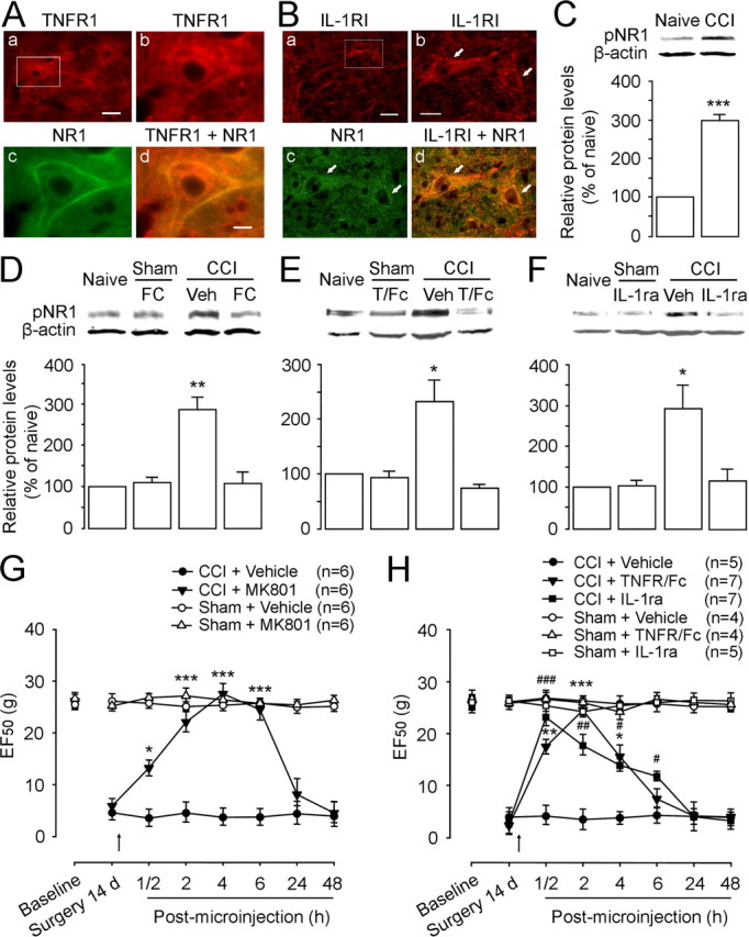
Spinal glial reaction and proinflammatory cytokine induction play an important role in the development of chronic pain states after tissue and nerve injury. The present study investigated the cellular and molecular mechanisms underlying descending facilitation of neuropathic pain with an emphasis on supraspinal glial-neuronal relationships. An early and transient reaction of microglia and prolonged reaction of astrocytes were found after chronic constriction injury (CCI) of the rat infraorbital nerve in the rostral ventromedial medulla (RVM), a major component of brainstem descending pain modulatory circuitry. There were prolonged elevations of cytokines tumor necrosis factor-alpha (TNF-alpha) and interleukin-1beta (IL-1beta) after CCI, and they were expressed in RVM astrocytes at 14 d after injury. Intra-RVM injection of microglial and astrocytic inhibitors attenuated mechanical hyperalgesia and allodynia at 3 and 14 d after CCI, respectively. Moreover, TNFR1 and IL-1R, receptors for TNF-alpha and IL-1beta, respectively, were expressed primarily in RVM neurons exhibiting immunoreactivity to the NMDA receptor (NMDAR) subunit NR1. CCI increased TNFR1 and IL-1R levels and NR1 phosphorylation in the RVM. Neutralization of endogenous TNF-alpha and IL-1beta in the RVM significantly reduced CCI-induced behavioral hypersensitivity and attenuated NR1 phosphorylation. Finally, intra-RVM administration of recombinant TNF-alpha or IL-1beta upregulated NR1 phosphorylation and caused a reversible and NMDAR-dependent allodynia in normal rats, further suggesting that TNF-alpha and IL-1beta couple glial hyperactivation with NMDAR function. These studies have addressed a novel contribution of supraspinal astrocytes and associated cytokines as well as central glial-neuronal interactions to the enhancement of descending facilitation of neuropathic pain.


Figures

Figure 1.
Figure 1.
Mechanical hyperalgesia/allodynia and time course-dependent hyperactivation of astrocytes in the RVM after CCI-ION. A, Examples of S–R function curves illustrating the intensity-dependent head withdrawal responses to mechanical stimuli. There is a significant leftward shift of the curve at 1 d (green) and 14 d (red) after CCI (p < 0.001; n = 9 rats) compared with the pre-CCI baseline (blue). The leftward shift of the S–R curves indicates an increased response to suprathreshold stimuli and a decrease in EF50 value, suggesting the presence of mechanical hyperalgesia and allodynia. B, Time course of mechanical hyperalgesia and allodynia as indicated by a reduction of the EF50 values. CCI-ION results in mechanical hyperalgesia and allodynia that persists over the 28 d observation period. The rats receiving a sham operation show a moderate reduction of the EF50 values that lasted for 3 d, which is totally blocked by local tissue anesthesia with injection of 0.25% bupivacaine (twice at 1–3 d) at the intraoral incision site in sham-operated rats. CCI-ION versus sham, ***p < 0.001; sham versus naive, #p < 0.05. C, Immunohistochemistry of GFAP in the RVM. a, Low power of a tissue section at the rostral medulla level. b, Enlarged RVM region corresponding to the small rectangle area in a. Enhanced expression of GFAP is observed at 3 d (c) and 14 d (d) after CCI, compared with GFAP immunostaining in naive animal (b). D, Immunostaining of S100β in the RVM. Increased S100β expression is observed at 3 d (b), 14 d (c), and 28 d (d) after CCI, compared with naive (a). E, Western blots illustrating CCI-induced increase in GFAP in the tissues punched out from the RVM. A representative blot is shown on top, and the mean protein levels are summarized below. Compared with naive and sham-operated (−) rats, the level of GFAP is selectively increased at the 14 and 28 d time points in CCI-ION (+) rats. The increase in GFAP at 1–3 d in sham-operated and CCI-ION rats is consistent with behavioral changes at these time points (B). β-Actin was used as a loading control. The asterisks indicate significant differences from the naive rats (*p < 0.05; n = 3 per time points). F, Western blots illustrating the effect of bupivacaine, a long-lasting local anesthetic, on the incision-induced increase in GFAP expression in the RVM at 3 d after sham operation. Bupivacaine (0.25%; 0.1 ml) is injected at the intraoral incision site twice in the first 3 d (+). Saline is injected as a vehicle control (−). Compared with naive rats, the GFAP expression is increased at 3 d after sham operation (*p < 0.05; n = 3 per group), which is completely abolished by the bupivacaine (n = 3). Scale bars: Ca, 0.5 mm; Cd (for Cb–d), Dd (for Da–d), 0.025 mm. NGC, Nucleus reticularis gigantocellularis; Py, pyramidal tract. Error bars indicate SEM.
Figure 2.
Figure 2.
Time course-dependent hyperactivation of microglia in the RVM after CCI. A, CD11b immunostaining in the RVM. a, Low power of a section at the rostral medulla level. b, Enlarged RVM region corresponding to the small rectangle area in a. CD11b expression is enhanced at 3 d (c) but not 14 d (d) after CCI-ION, compared with CD11b immunoreactivity in naive animal (b). B, Iba1 immunostaining in the RVM. Enhanced Iba1 expression is observed at 3 d (b) and declined to near control expression levels at 14 d (c) and 28 d (d) after CCI, compared with naive animal (a). C, Western blots illustrating CCI-induced short-lasting increase in CD11b in RVM tissues. Compared with naive rats, CD11b expression is temporarily increased at the 1–3 d time points but not at 14 and 28 d in both sham-operated (−) and CCI-ION (+) rats. The asterisks indicate significant differences from the naive rats (*p < 0.05; n = 3 per time points). D, Local tissue anesthesia (+), compared with saline injection (−), completely blocks sham operation-induced increase in CD11b in the RVM at 3 d, when compared with naive rats [sham (−) vs naive, p < 0.05; n = 3 per group]. Scale bars: Aa, 0.5 mm; Ad (for Ab–d), Bd (for Ba–d), 0.025 mm. NGC, Nucleus reticularis gigantocellularis. Error bars indicate SEM.
Figure 3.
Figure 3.
Western blot analysis shows inhibition of fluorocitrate on glial hyperactivation and functions. A, B, Microinjection of fluorocitrate (FC+; 100 pmol) significantly inhibits enhanced expression of GFAP (A) and S100β (B) at 14 d after CCI-ION (p < 0.05; n = 3 per group) but does not affect their expression at 14 d after sham operation, compared with vehicle treatment (−). C, D, Fluorocitrate (100 pmol) significantly reverses upregulation of TNF-α (C) and IL-1β (D) at 14 d after CCI-ION (p < 0.05; n = 3 per group) but does not change their expression at 14 d after sham operation, compared with vehicle treatment (−). E, There are no effects of FC (100 pmol) on increases in CD11b expression at 3 d after sham operation and CCI-ION, compared with vehicle (Veh) (n = 3 per group). However, the microglial inhibitor MC (1 pmol) attenuates or abolishes CD11b increase in the RVM at 3 d after sham or CCI treatment. All asterisks indicate p < 0.05 when compared with the naive (n = 3 per group) in A–E. Error bars indicate SEM.
Figure 4.
Figure 4.
The different effect of glial inhibitors (propentofylline, fluorocitrate, and minocycline) on behavioral hyperalgesia/allodynia at early (3 d) and later (14 d) time points after CCI-ION. At posttreatment, the glial inhibitor propentofylline (A), the selective astrocytic inhibitor fluorocitrate (B), and the microglial inhibitor minocycline (C) are microinjected into the RVM at 3 d (right column) and 14 d (left column) after sham operation or CCI-ION. The dose-dependent effects of the glial inhibitors on behavioral hyperalgesia/allodynia are measured at 14 d after the surgery. An effective high dose of the inhibitor is then used for behavioral observation at the 3 d time point. Vehicles are microinjected as a control. A, PPF (1 fmol, 100 fmol, and 10 pmol) produces a dose-dependent attenuation of CCI-induced mechanical hyperalgesia and allodynia at 14 d, persistent for 4–24 h compared with vehicle microinjection in CCI rats (left). High-dose propentofylline (10 pmol) has no effect on EF50 values at 14 d in sham rats (left). This dose of propentofylline transiently blocks moderate hyperalgesia and allodynia at 3 d compared with vehicle in sham-operated rats (right), and also significantly abolishes mechanical allodynia at 3 d after CCI compared with vehicle in CCI rats (right). B, Two doses of FC (1 and 100 fmol) after microinjection into the RVM significantly attenuate hyperalgesia and allodynia similarly at least for 6 h, compared with vehicle treatment at 14 d in CCI rats (left). High dose of fluorocitrate (100 fmol) has no effect on EF50 values at 14 d in sham rats (left). However, fluorocitrate (100 fmol) does not prevent CCI-induced behavioral hypersensitivity to mechanical stimulation at 3 d after CCI or sham operation (right). C, In contrast, microinjection of MC (1 pmol) into the RVM significantly reduces hyperalgesia and allodynia for 6 h at 3 d after CCI-ION compared with vehicle treatment in CCI rats, and also transiently blocks sham operation-induced mechanical hypersensitivity (right). Lower dose of minocycline (10 fmol) induced a slight and short-lasting inhibition for CCI-induced allodynia (right). The two doses of minocycline have no effect on CCI-induced hyperalgesia and allodynia at 14 d (left). Also, there are no changes in EF50 values after minocycline (1 pmol) injection at 14 d after sham-operated rats. ***, ###p < 0.001; **p < 0.01; *, #,^ p < 0.05 versus CCI plus vehicle in left. ***p < 0.001, *p < 0.05, CCI plus drug versus CCI plus vehicle in right. #p < 0.05, sham plus drug versus sham plus vehicle in right. Error bars indicate SEM.
Figure 5.
Figure 5.
Enhanced expression of TNF-α and IL-1β in the RVM at 14 d after CCI-ION. A, Western blots illustrating that the levels of TNF-α (left) and IL-1β (right) are increased in the RVM in CCI-ION rats compared with sham-operated rats (*p < 0.05; n = 3 per group). The anti-TNF-α antibody identifies both the transmembrane TNF-α precursor (26 kDa) and matured TNF-α (17 kDa). Both TNF-α precursor and matured TNF-α exhibit an increase in the RVM at 14 d after CCI-ION. Error bars indicate SEM. B, Immunostaining of TNF-α and IL-1β in RVM. a, d, Low power of a tissue section at the rostral medulla level. b, e, Enlarged RVM region corresponds to the rectangle area in a and d, respectively. Compared with naive rats (b, e), there is an increase in TNF-α IR and IL-1β IR in RVM at 14 d after CCI-ION (c, f). The arrowheads indicate positively labeled cells. C, RVM tissue sections at 14 d after CCI are double stained for TNF-α (a, d; red) and GFAP (b; green) or NeuN (e; green). The arrowheads indicate positively labeled cells. Overlap of a and b reveals that the TNF-α IR is colocalized with GFAP IR in RVM cells (c; yellow, arrows), suggesting its presence in astrocytes. Overlap of d and e shows a lack of TNF-α in RVM neurons (f). D, RVM tissue sections were double stained for IL-1β (a, d; red) and GFAP (b; green) or NeuN (e; green). The arrowheads indicate positively labeled cells. Overlap of a and b reveals that the IL-1β-labeled cells are GFAP immunoreactive in RVM (c; yellow, arrows), suggesting its presence in astrocytes. Overlap of d and e shows a lack of IL-1β IR in these RVM neurons (f). Scale bars: Ba,d, 0.1 mm; Bb,c,e,f, 0.03 mm; C, D, 0.03 mm.
Figure 6.
Figure 6.
Enhanced expression of cytokine receptors for TNF-α and IL-1β in RVM neurons at 14 d after CCI-ION. A, C, Immunostaining shows distribution of TNFR1 (A; red) and IL-1RI expression (B; red) in low power of a tissue section at the rostral medulla level (a) and enlarged RVM region (b) corresponding to the small rectangle area in a. The arrowheads in b and c indicate single-labeled neurons. Double labeling shows TNFR1-labeled (Ab) and IL-1RI-labeled cells (Cb) are immunoreactive to NeuN (Ac, Cc; green), suggesting their expression in the RVM neurons (Ad, Cd; arrows). B, D, Western blots illustrating that the levels of TNFR1 (B) and IL-1RI (D) are increased in the RVM tissues at 14 d in CCI-ION rats compared with sham-operated rats (p < 0.05; n = 3 per group). *p < 0.05 versus naive in B and D. Scale bars: Aa, Ca, 0.1 mm; Ab–d, Cb–d, 0.03 mm. Error bars indicate SEM.
Figure 7.
Figure 7.
The effect of cytokine inhibitors on NMDAR subunit NR1 phosphorylation in the RVM and behavioral hyperalgesia and allodynia at 14 d after CCI-ION. A, B, Double immunostaining shows colocalization of TNFR1 (Ab; red) or IL-1RI (Bb; red) with NR1 (Ac, Bc; green) in RVM neurons. Overlay of b and c reveals double labeling of RVM neurons with TNFR1/NR1 (Ad; yellow-orange) or IL-1RI/NR1 (Bd; yellow-orange). Note colocalization of these receptors with NR1 in the cell membrane. C, Compared with naive rats, the level of pNR1ser896 is increased at 14 d after CCI-ION (p < 0.001; n = 3 per group). D, The increased pNR1ser896 is totally blocked by intra-RVM microinjection of astrocytic inhibitor FC (100 fmol) compared with naive and sham-operated rats (n = 3 per group). E, F, Neutralizing endogenous TNF-α and IL-1β in the RVM using TNFR1/Fc (T/Fc) (50 fmol) (E) and IL-1ra (3 pmol) (F), respectively, blocks CCI-induced increase in pNR1 expression in the RVM at 14 d after CCI-ION compared with sham treatment (p < 0.05; n = 3 per group). No effect of these inhibitors on basal pNR1 expression is observed in sham-operated rats compared with naive rats (n = 3). G, Microinjection of NMDAR channel blocker MK801 (10 pmol) into the RVM abolishes CCI-induced mechanical hyperalgesia and allodynia compared with vehicle (Veh) injection at 14 d after CCI-ION. H, No effect of intra-RVM TNFR1/Fc (50 fmol) and IL-1ra (3 pmol) on basal mechanical threshold is observed in sham-operated rats compared with naive rats. However, these doses of TNFR1/Fc and IL-1ra attenuate CCI-ION-induced hyperalgesia/allodynia for 4–6 h after microinjection compared with vehicle treatment in CCI rats. D–F, **p < 0.01; *p < 0.05 versus naive; G, H, ***, ###p < 0.001; ** , ##p < 0.01; *, #p < 0.05 versus CCI plus vehicle. Scale bars: Aa, Ba, 0.015 mm; Ab–d, Bb–d, 0.005 mm. Error bars indicate SEM.
Figure 8.
Figure 8.
Intra-RVM rTNF-α and rIL-1β enhance pNR1 levels and produce descending pain facilitation in normal rats. A, Microinjection of rTNF-α (120 fmol) and rIL-1β (120 fmol) into the RVM enhances expression of pNR1ser896 in the RVM tissue compared with baseline levels in naive rats (p < 0.05; n = 4 per group). Pretreatment of an NMDAR channel blocker MK801 (+) (n = 4) completely prevents rTNF-α- and rIL-1β-induced increase in pNR1 compared with vehicle treatment (−). RVM tissues are collected at 2 h after microinjection of the recombinant agents into the RVM. B, Mechanical sensitivity of the skin is assessed after microinjection of rTNF-α. The behavioral hyperalgesia and allodynia develops and lasts for 4 h after microinjection of rTNF-α (120 fmol) as indicated by a reduction of EF50 values (p < 0.05). Pretreatment of MK801 (10 pmol) significantly abolishes rTNF-α-produced mechanical hypersensitivity compared with vehicle plus rTNF-α. C, Microinjection of rIL-1β (120 fmol) results in a significant reduction of EF50 values for 2 h (p < 0.05), which is prevented by pretreatment with MK801. *p < 0.05, rTNF-α or rIL-1β versus vehicle injection. Error bars indicate SEM.

Similar articles

Cited by

References

    1. Apkarian AV, Lavarello S, Randolf A, Berra HH, Chialvo DR, Besedovsky HO, del Rey A. Expression of IL-1β in supraspinal brain regions in rats with neuropathic pain. Neurosci Lett. 2006;407:176–181. - PMC - PubMed
    1. Beattie EC, Stellwagen D, Morishita W, Bresnahan JC, Ha BK, Von Zastrow M, Beattie MS, Malenka RC. Control of synaptic strength by glial TNFalpha. Science. 2002;295:2282–2285. - PubMed
    1. Bennett GJ, Xie YK. A peripheral mononeuropathy in rat that produces abnormal pain sensation like those seen in man. Pain. 1988;33:87–107. - PubMed
    1. Burgess SE, Gardell LR, Ossipov MH, Malan TP, Jr, Vanderah TW, Lai J, Porreca F. Time-dependent descending facilitation from the rostral ventromedial medulla maintains, but does not initiate, neuropathic pain. J Neurosci. 2002;22:5129–5136. - PMC - PubMed
    1. Bursztajn S, Rutkowski MD, Deleo JA. The role of the N-methyl-d-aspartate receptor NR1 subunit in peripheral nerve injury-induced mechanical allodynia, glial activation and chemokine expression in the mouse. Neuroscience. 2004;125:269–275. - PubMed

Publication types

MeSH terms