. 2008 Oct 15;22(20):2767-72.
doi: 10.1101/gad.503108.

DNA helicases Sgs1 and BLM promote DNA double-strand break resection

Affiliations

DNA helicases Sgs1 and BLM promote DNA double-strand break resection

Serge Gravel et al. Genes Dev. .

Abstract

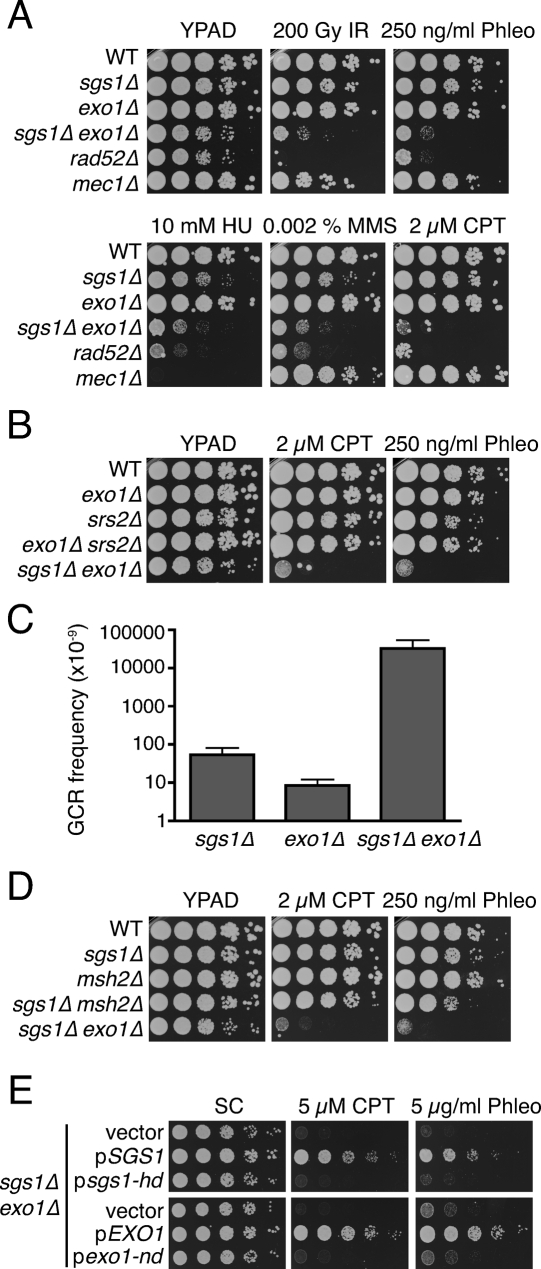
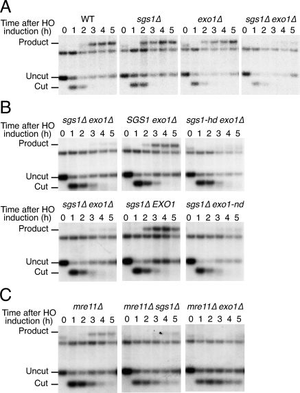
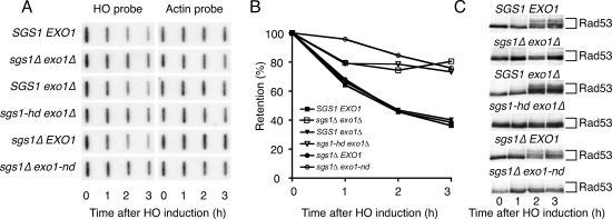
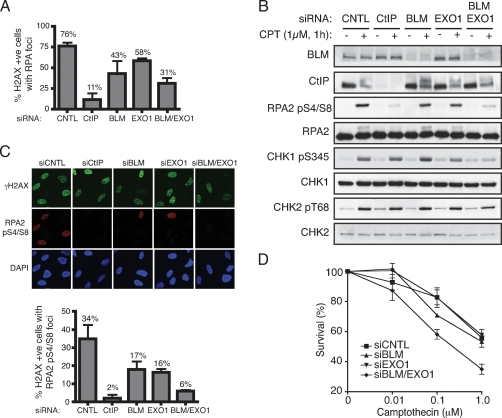
A key cellular response to DNA double-strand breaks (DSBs) is 5'-to-3' DSB resection by nucleases to generate regions of ssDNA that then trigger cell cycle checkpoint signaling and DSB repair by homologous recombination (HR). Here, we reveal that in the absence of exonuclease Exo1 activity, deletion or mutation of the Saccharomyces cerevisiae RecQ-family helicase, Sgs1, causes pronounced hypersensitivity to DSB-inducing agents. Moreover, we establish that this reflects severely compromised DSB resection, deficient DNA damage signaling, and strongly impaired HR-mediated repair. Furthermore, we show that the mammalian Sgs1 ortholog, BLM--whose deficiency causes cancer predisposition and infertility in people--also functions in parallel with Exo1 to promote DSB resection, DSB signaling and resistance to DSB-generating agents. Collectively, these data establish evolutionarily conserved roles for the BLM and Sgs1 helicases in DSB processing, signaling, and repair.


Figures

Figure 1.
Figure 1.
SGS1 and EXO1 are components of parallel pathways promoting resistance to DNA-damaging agents. (A) sgs1Δ exo1Δ double mutants are hypersensitive to DNA damaging agents. Tenfold serial dilutions of the indicated strains were treated with IR or were plated on media containing the indicated drug, then were incubated for 3 d at 30°C. (B) Srs2 is not required for resistance to DNA damaging agents in the absence of Exo1. Strains were treated as in A. (C) GCR frequency was measured for the indicated strains; the mean and standard deviation of two fluctuation tests are shown. (D) Inactivation of MMR does not sensitize sgs1Δ cells to DNA damaging agents. Analyses were as in A. (E) The catalytic activities of Sgs1 and Exo1 promote resistance to DNA damaging agents. sgs1Δ exo1Δ mutants were transformed with an empty vector, with a vector expressing the wild-type (pSGS1) or a helicase-deficient (psgs1-hd) version of Sgs1, or with a vector expressing the wild-type (pEXO1) or a nuclease-deficient (pexo1-nd) version of Exo1. Analyses were as in A.
Figure 2.
Figure 2.
sgs1Δ exo1Δ mutants have mating-type switching defects. (A) Time course of DSB repair at the MAT locus in the indicated strains. Galactose was added at time 0 to induce HO expression, and samples were removed at 1-h intervals for Southern blot analysis. Bands corresponding to uncut (MATa), HO endonuclease-cut and product (MATα) restriction fragments are indicated. The unlabeled band corresponds to the distal fragment and serves as a loading control. (B) The catalytic activities of Sgs1 and Exo1 promote mating-type switching. sgs1Δ exo1Δ cells transformed with an empty vector, or with a vector expressing the wild-type (pSGS1) or a helicase-deficient (psgs1-hd) version of Sgs1 (top panel), or with a vector expressing the wild-type (pEXO1) or a nuclease-deficient (pexo1-nd) version of Exo1 (bottom panel) were analyzed as in A. (C) Mre11 and Sgs1 promote mating-type switching by independent pathways. Analyses were as in A.
Figure 3.
Figure 3.
Sgs1 and Exo1 act in parallel to promote DSB resection and checkpoint activation. (A) sgs1Δ exo1Δ mutants have resection defects. sgs1Δ exo1Δ cells containing an irreparable HO site were transformed with plasmids to generate the indicated strains. Galactose was added to cultures at time 0 and samples were removed at 1-h intervals for DNA analysis by denaturing slot blotting with a probe specific to sequences adjacent to the HO site. The blot was then stripped and reprobed with actin as a loading control. (B) Densitometric analysis of the data in A. For each time point, the signal detected by the HO probe was normalized to actin. (C) Extracts from samples taken during the experiment shown in A were analyzed by Western blotting with an anti-Rad53 antibody.
Figure 4.
Figure 4.
BLM and EXO1 promote DNA DSB resection and associated events in human cells. (A) BLM and EXO1 deficiency impairs Camptothecin-induced RPA focus formation. U2OS cells were transfected with siRNAs directed against Luciferase (siCNTL), CtIP, BLM, EXO1, or a combination of BLM and EXO1, then 72 h later were mock-treated or treated with 1 μM Camptothecin for 1 h. Cells were next detergent-extracted and fixed, then foci for phosphorylated histone H2AX (γH2AX) and RPA2 were visualized by indirect immunofluorescence. More than 100 cells were counted for each sample and the percentages of cells exhibiting both γH2AX and RPA foci was determined. Data represent the mean ± SEM from three independent experiments. All counting was done blind. (B) BLM and EXO1 promote DSB signaling. Extracts of mock-treated or Camptothecin-treated cells depleted for the indicated factors were analyzed by Western blotting with the indicated antibodies. Endogenous EXO1 levels were too low to allow detection with anti-EXO1 antisera and so verification of EXO1 siRNA depletion was done with cells stably expressing a GFP-Exo1 construct (see Supplemental Fig. S4). (C) RPA Ser-4 and Ser-8 phosphorylation (RPApS4/S8) is compromised by BLM and EXO1 depletion. CtIP, BLM, or EXO1 were depleted and cells were treated as in A, followed by analysis by indirect immunofluorescence with the indicated antibodies. More than 100 cells were counted for each sample, and data represent the mean ± SEM from three independent experiments. Counting was done blind. (D) Codepletion or BLM and EXO1 yields Camptothecin hypersensitivity. Seventy-two hours following transfection with the indicated siRNAs, U2OS cells were treated with Camptothecin for 1 h, and cell survival was determined by colony formation. Data represent the mean ± SEM from three independent experiments.

Comment in

Similar articles

Cited by

References

    1. Anantha R.W., Vassin V.M., Borowiec J.A. Sequential and synergistic modification of human RPA stimulates chromosomal DNA repair. J. Biol. Chem. 2007;282:35910–35923. - PubMed
    1. Bachrati C.Z., Hickson I.D. RecQ helicases: Suppressors of tumorigenesis and premature aging. Biochem. J. 2003;374:577–606. - PMC - PubMed
    1. Bermejo R., Doksani Y., Capra T., Katou Y.M., Tanaka H., Shirahige K., Foiani M. Top1- and Top2-mediated topological transitions at replication forks ensure fork progression and stability and prevent DNA damage checkpoint activation. Genes & Dev. 2007;21:1921–1936. - PMC - PubMed
    1. Clerici M., Mantiero D., Lucchini G., Longhese M.P. The Saccharomyces cerevisiae Sae2 protein negatively regulates DNA damage checkpoint signalling. EMBO Rep. 2006;7:212–218. - PMC - PubMed
    1. Cobb J.A., Bjergbaek L., Gasser S.M. RecQ helicases: At the heart of genetic stability. FEBS Lett. 2002;529:43–48. - PubMed

Publication types

MeSH terms