. 2008 Oct 15;22(20):2843-55.
doi: 10.1101/gad.1708608.

RMI, a new OB-fold complex essential for Bloom syndrome protein to maintain genome stability

Affiliations

RMI, a new OB-fold complex essential for Bloom syndrome protein to maintain genome stability

Dongyi Xu et al. Genes Dev. .

Abstract

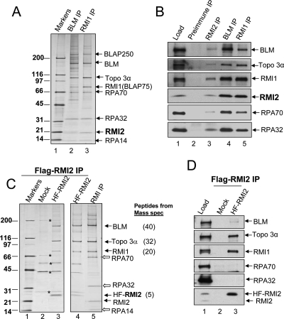
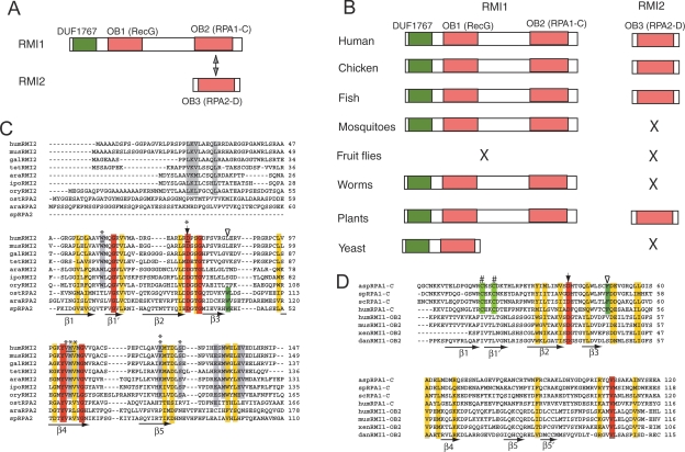
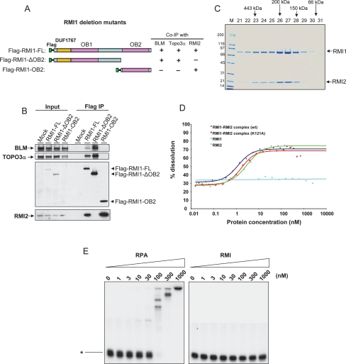
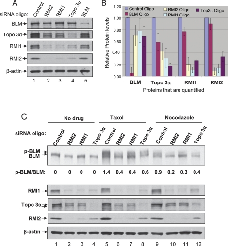
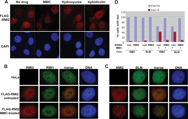
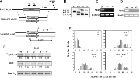
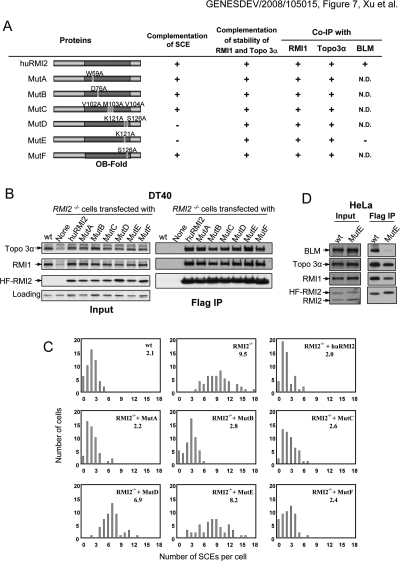
BLM, the helicase mutated in Bloom syndrome, associates with topoisomerase 3alpha, RMI1 (RecQ-mediated genome instability), and RPA, to form a complex essential for the maintenance of genome stability. Here we report a novel component of the BLM complex, RMI2, which interacts with RMI1 through two oligonucleotide-binding (OB)-fold domains similar to those in RPA. The resulting complex, named RMI, differs from RPA in that it lacks obvious DNA-binding activity. Nevertheless, RMI stimulates the dissolution of a homologous recombination intermediate in vitro and is essential for the stability, localization, and function of the BLM complex in vivo. Notably, inactivation of RMI2 in chicken DT40 cells results in an increased level of sister chromatid exchange (SCE)--the hallmark feature of Bloom syndrome cells. Epistasis analysis revealed that RMI2 and BLM suppress SCE within the same pathway. A point mutation in the OB domain of RMI2 disrupts the association between BLM and the rest of the complex, and abrogates the ability of RMI2 to suppress elevated SCE. Our data suggest that multi-OB-fold complexes mediate two modes of BLM action: via RPA-mediated protein-DNA interaction, and via RMI-mediated protein-protein interactions.


Figures

Figure 1.
Figure 1.
RMI2 is an integral component of BLM complexes. (A) A silver-stained SDS gel showing the presence of RMI2 in complexes immunoprecipitated by both BLM and RMI1 antibodies. The identity of different BLM components has been determined previously by mass spectrometry and immunoblotting (also see B). (B) Immunoblotting to show that RMI2 coimmunoprecipitates with other components of BLM complexes. Immunoprecipitation (IP) with preimmune serum was included as a negative control. The nuclear extract input (Load) is also shown. (C,D) A silver-stained SDS gel (C) and immunoblotting (D) show that 6-histidine and Flag double-tagged RMI2 (HF-RMI2) coimmunoprecipitated with BLM, Topo 3α, and RΜΙ1, but not RPA. The major polypeptides corresponding to the BLM components identified by mass spectrometry, and the numbers of peptides identified are indicated on the right of the figure. As a control, mock IP (Mock) was performed by using regular HeLa cells that do not express HF-RMI2. Several contaminating polypeptides are marked by asterisks. IP was performed using Flag antibody. We failed to detect RPA in the BLM complex isolated using the HF–RMI2 antibody (C,D), even though RPA was detectable in the complex immunoprecipitated using the antibody against endogenous RMI2 (B). One possibility is that the existence of the HF tag interferes with the interaction between HF-RMI2 and RPA.
Figure 2.
Figure 2.
hRMI2 and hRMI1 contain three putative OB domains, two of which resemble a pair of interacting domains in RPA. (A) Schematic representation of hRMI1 and hRMI2. RMI1 contains two OB-fold domains: OB1 is most similar to the Wedge domain in bacterial RecG helicases (Supplemental Fig. 2), whereas OB2 resembles the RPA1-C domain (see D). RMI2 has one OB domain (OB3), which resembles that of RPA2-D (see C). DUF1767 is a domain of unknown function, but is conserved in RMI1 in all species. (B) Schematic representation of RMI1 and RMI2 in different eukaryotic species. The absence of an obvious ortholog is indicated by “X.” The orthologs were identified by using the BLASTP algorithm to search the NR database maintained at NCBI. The orthologs include chicken RMI1 (NP_001026783) and RMI2 (NP_001006174.1); zebrafish RMI1 (AAH45482) and RMI2 (XP_691674.1); mosquito RMI1 (Anopheles gambiae XP_552585); worm RMI1 (C. elegans NP_741607); plant RMI1 (Arabidopsis thaliana NP_201159) and RMI2 (NP_172315.1); budding yeast RMI1 (Q02685). (C,D) Sequence alignment of RMI2 and RPA2 (C) or RMI1-OB2 and RPA1-C (D). The five predicted β-strands of the OB-folds are underlined with arrows. The identical and conserved residues are highlighted with red and yellow, respectively. Gray highlighted residues are only conserved in RMI2. Solid arrows indicate a highly conserved aspartic acid residue. Green highlighting and empty arrowheads mark the conserved aromatic residues conserved in RPA but not RMI. The residues mutated in Figure 7 are marked with an asterisk. K121, the mutation of which disrupts RMI2 function, is indicated by two asterisks. Two cysteine residues that are part of the Zn-finger in RPA1-C are marked with “#,” but these are not conserved in RMI1-OB2. The abbreviations and gene access numbers are as follows. RMI2: (mus) mouse, Q3UPE3; (gal) chicken; (tet) Tetraodon nigroviviridis, CAG1226.1; (ara) Arabidopsis thaliana, NP_172315.1; (ipo) Ipomoea trifida, BAF36320.1; (ory) Oryza sativa, AAK14410.1. RPA2: (ost) Ostreococcus tauri, CAL56444.1; (ara) Arabidopsis, NP_565571; (sp) Schizosaccharomyces pombe, NP_588227.2. RPA1: (asp) Aspergillus, XP_00127599.1; (sp) S. pombe, NP_595092.1; (sc) Saccharyomyces cerevisiae, NP_009404.1. RMI1: (xen) Xenopus laevis; (dan) Danio rerio.
Figure 3.
Figure 3.
RMI2 and RMI1 constitute a new type of OB-fold complex that lacks detectable ssDNA-binding activity but is capable of stimulating dHJ dissolution. (A) Schematic representation of different RMI1 deletion mutants (left), and their ability to coimmunoprecipitate with different BLM complex components from HeLa extract (right). (B) IP-Western showing that RMI1 deletion mutants in A coimmunoprecipitate with different BLM complex components. The Flag-tagged full-length (FL) or different deletion mutants of RMI1 were transfected into HeLa cells, and coimmunoprecipitation was performed using the Flag antibody. (C) A Coomassie-stained SDS-gel shows the purified recombinant RMI1–RMI2 complex fractionated by Sephadex 200 gel filtration column. RMI1 was fused with a 6xhistidine tag, whereas RMI2 was fused with a strep tag. The proteins were coexpressed in E. coli and were purified as described in Supplemental Material. Proteins with known molecular mass were used to calibrate the column, and their elution positions are shown above the figure. Notably, the peaks of the two RMI proteins are coincident, indicating that they are present in the same complex. (D) A graph showing stimulation of dHJ dissolution by the recombinant RMI complex containing wild-type RMI1–RMI2 (wt), RMI1–RMI2 (K121A) mutant, RMI1 purified from yeast, and RMI2. All reactions contained Topo 3α (22.5 nM) and BLM (10 nM). The primary image data of dissolution are shown in Supplemental Figure 3. (E) Gel-shift DNA-binding assay to show that the RMI complex exhibits little or no detectable ssDNA-binding activity (right panel) compared with RPA (left panel). Recombinant RPA complex was purified from E. coli and kindly provided by Dr. Guomin Li. A p32-labeled 94-nucleotide ssDNA is indicated on the left. The concentrations of different proteins are shown on the top.
Figure 4.
Figure 4.
The RMI1–RMI2 complex is required for BLM complex stability and mitotic phosphorylation of BLM. (A,B) Immunoblotting (A), and its quantification (B), to show that HeLa cells depleted of RMI2, RMI1, or Topo 3α, exhibited reduced levels of other BLM complex components. Immunoblotting for β-actin was included as a loading control. In B, at least four independent siRNA depletion experiments have been performed for each protein, and the immunoblotting data were quantified using TotalLab software. The protein level in cells treated with control siRNA was designated as 1. Data represent the mean values, and the bars represent standard error. (C) Immunoblotting shows that mitotic phosphorylation of BLM is reduced in cells depleted of RMI2, RMI1, or Topo 3α. Cells were either untreated (no drug), or treated with mitosis inhibitors Taxol (1 μM) or Nocodazole (100 ng/mL) for 16 h, as shown above the lanes. The hyperphosphorylated BLM is indicated (p-BLM). The ratio between hyper- and hypophosphorylated BLM was quantified using image software, and is shown below the image.
Figure 5.
Figure 5.
RMI2 colocalizes with BLM and RMI1 in nuclear foci in response to DNA damage, and is required for focus formation. (A) Immunofluorescence showing that RMI2 is redistributed to nuclear foci in response to several DNA damaging agents, including MMC, hydroxyurea, and aphidicolin. (B,C) Immunofluorescence showing that Flag-tagged RMI2 (Flag-RMI2) colocalizes with RMI1 (B) and BLM (C) in nuclear foci in response to MMC treatment. HeLa cells that are not transfected with Flag-RMI2 were included as a negative control. (D) A histogram to illustrate that the percentage of cells containing BLM or RMI1 foci is drastically reduced in HeLa cells depleted of RMI2 in response to MMC treatment.
Figure 6.
Figure 6.
RMI2-null DT40 cells resemble BLM mutant cells in displaying an elevated level of SCE. (A) Schematic representation of the chicken RMI2 genomic DNA, the two targeting vectors, and the restriction map of the locus after knockout. Two restriction enzyme digestion sites used in Southern Blotting, XhoI and SalI, are marked as X and S, respectively. The primers used for genomic-PCR analysis (P3–P2) are depicted in B. Southern blotting shows that both alleles of the RMI2 gene have been inactivated by the targeting vectors described in A. The genomic DNA from wild-type (wt), RMI2+/−, and RMI2−/− cells was first digested with XhoI and SalI restriction enzymes, and subsequently hybridized with the probe shown in A. (C) Genomic-PCR analysis to show the absence of RMI2 genomic DNA in RMI2−/− cells. The FANCC gene was included as a positive control. (D) RT–PCR analysis to show that RMI2 mRNA is undetectable in RMI2−/− cells. RAD51 was included as a positive control. (E) Immunoblotting to show that Topo 3α and RMI1 levels are reduced in four different clones of RMI2−/− cells. (F) Histograms showing the SCE levels of wild-type, RMI2−/− cells, BLM−/−, and RMI2−/−/BLM−/− cells. The mean number of SCEs per cell is shown in the top right corner.
Figure 7.
Figure 7.
An RMI2 point mutant defective in mediating interactions with BLM is deficient in suppressing SCE in RMI2 mutant cells. (A) Schematic representation of different hRMI2 point mutants and their ability to suppress the elevated SCE level (C) and restore the protein stability of RMI1 and Topo 3α (B) in RMI2−/− DT40 cells. Their ability to interact with BLM complex components based on B and D are also shown. The amino acid residues mutated are shown in Figure 2C. (N.D) Not determined. (B, left panels) Immunoblotting showing that human wild-type and different point mutants of RMI2 can complement the phenotype of reduced stability of Topo 3α and RMI1 of RMI2−/− cells. (Right panels) Moreover, these point mutants can efficiently coimmunoprecipitate with Topo 3α and RMI1, indicating that their ability to interact with these two proteins remains largely intact. The wild-type and mutant RMI2 are all fused to a Flag-epitope, and immunoprecipitation was performed using the Flag antibody. (C) Histograms showing SCE levels of RMI2−/− DT40 cells complemented by wild-type and various point mutants of hRMI2 as illustrated in A. The mean number of SCEs per cell is shown in the top right corner. (D) Immunoblotting shows that the K121A mutant (MutE) coimmunoprecipitated with drastically reduced level of BLM, indicating that this mutant is defective in interacting with BLM.

Comment in

References

    1. Bachrati C.Z., Hickson I.D. RecQ helicases: Guardian angels of the DNA replication fork. Chromosoma. 2008;117:219–233. - PubMed
    1. Bachrati C.Z., Borts R.H., Hickson I.D. Mobile D-loops are a preferred substrate for the Bloom’s syndrome helicase. Nucleic Acids Res. 2006;34:2269–2279. - PMC - PubMed
    1. Bischof O., Kim S.H., Irving J., Beresten S., Ellis N.A., Campisi J. Regulation and localization of the Bloom syndrome protein in response to DNA damage. J. Cell Biol. 2001;153:367–380. - PMC - PubMed
    1. Bochkareva E., Korolev S., Bochkarev A. The role for zinc in replication protein A. J. Biol. Chem. 2000;275:27332–27338. - PubMed
    1. Briggs G.S., Mahdi A.A., Wen Q., Lloyd R.G. DNA binding by the substrate specificity (wedge) domain of RecG helicase suggests a role in processivity. J. Biol. Chem. 2005;280:13921–13927. - PubMed

Publication types

MeSH terms

LinkOut - more resources