. 2008 Dec;19(12):5398-408.
doi: 10.1091/mbc.e08-07-0704. Epub 2008 Oct 15.

Analysis of prelamin A biogenesis reveals the nucleus to be a CaaX processing compartment

Affiliations

Analysis of prelamin A biogenesis reveals the nucleus to be a CaaX processing compartment

Jemima Barrowman et al. Mol Biol Cell. 2008 Dec.

Abstract

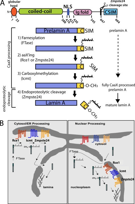
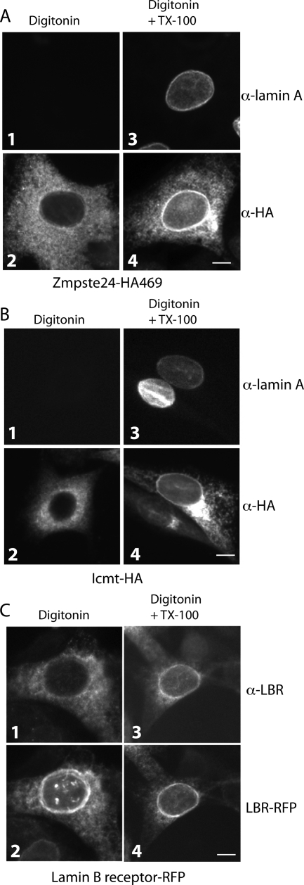
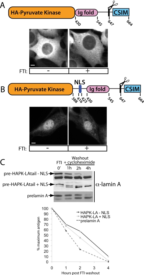
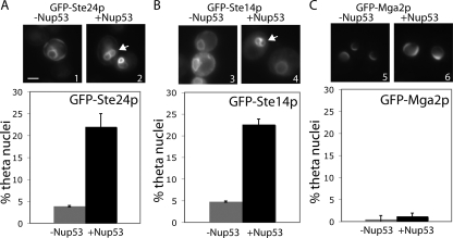
Proteins establish and maintain a distinct intracellular localization by means of targeting, retention, and retrieval signals, ensuring most proteins reside predominantly in one cellular location. The enzymes involved in the maturation of lamin A present a challenge to this paradigm. Lamin A is first synthesized as a 74-kDa precursor, prelamin A, with a C-terminal CaaX motif and undergoes a series of posttranslational modifications including CaaX processing (farnesylation, aaX cleavage and carboxylmethylation), followed by endoproteolytic cleavage by Zmpste24. Failure to cleave prelamin A results in progeria and related premature aging disorders. Evidence suggests prelamin A is imported directly into the nucleus where it is processed. Paradoxically, the processing enzymes have been shown to reside in the cytosol (farnesyltransferase), or are ER membrane proteins (Zmpste24, Rce1, and Icmt) with their active sites facing the cytosol. Here we have reexamined the cellular site of prelamin A processing, and show that the mammalian and yeast processing enzymes Zmpste24 and Icmt exhibit a dual localization to the inner nuclear membrane, as well as the ER membrane. Our findings reveal the nucleus to be a physiologically relevant location for CaaX processing, and provide insight into the biology of a protein at the center of devastating progeroid diseases.


Figures

Figure 1.
Figure 1.
Schematics depicting the structure, steps and possible cellular sites of posttranslational processing of prelamin A. (A) Diagram showing the structural domains and corresponding amino acid numbers for human prelamin A (top). Processing steps and enzymes involved in the conversion of prelamin A to mature lamin A (bottom, see text for details). Steps 1–3 constitute CaaX-processing steps common to many proteins and are carried out by the enzymes indicated in parentheses, whereas step 4, mediated by Zmpste24, is an endoproteolytic cleavage event unique to prelamin A that cleaves the final 15 amino acids from prelamin A to yield mature lamin A. Note that the aaX'ing enzyme for prelamin A (either Rce1 or Zmpste24) has not yet been established. (B) Two opposing models for the intracellular location of prelamin A processing. Cytosol/ER processing (left) is supported by knowledge of CaaX enzyme localization to the cytosol (FTase) and ER membranes (Rce1, Icmt, Zmpste24) up to the time of this study. Nuclear processing (right) has been hypothesized based on lamin A localization and kinetic studies (Lehner et al., 1986; Goldman et al., 1992). Evidence for the presence of the processing enzymes on the INM is provided in the present study.
Figure 2.
Figure 2.
Prelamin A can be CaaX processed and endoproteolytically cleaved after preaccumulation within the nucleus. (A) An NIH 3T3 stable cell line was created to inducibly express GFP-prelamin A using the Clontech TetOff system. When expression is off, no signal is apparent. On induction (on), a nucleoplasmic and rim localization typical for lamin A is observed. When induced in the presence of farnesyltransferase inhibition (FTI), GFP-prelamin A accumulates within the nucleus in bright foci, as has also been documented by us and others (Capell et al., 2005; Glynn and Glover, 2005; Mallampalli et al., 2005; Toth et al., 2005; Yang et al., 2006). Bar, 50 μm. (B) Cycloheximide-chase analysis of preaccumulated nuclear prelamin A upon release from FTI block. Conversion of the GFP-prelamin A to GFP-lamin A corresponds with the disappearance of the prominent foci and appearance of GFP-lamin A at the nuclear rim. Bar, 5 μm. This experiment was performed three times with similar results, and a representative immunoblot is shown here. Quantitation of the above immunoblot reveals the half-life of maturation (disappearance of prelamin A and appearance of mature lamin A) to be approximately ∼100 min. (C) Maturation of nuclear-accumulated GFP-prelamin A is farnesylation dependent, as it does not occur for lamin A C661S. A C661S mutant, in which the CaaX motif cysteine has been mutated to serine, is not cleaved after nuclear preaccumulation and FTI washout (detected by anti-prelamin A antibody). Bar, 5 μm.
Figure 3.
Figure 3.
Heterokaryon fusion assay reveals prelamin A remains within the nucleus after FTI washout. (A) Schematic of the heterokaryon fusion assay used to examine the possibility of nuclear/cytosol shuttling of GFP-prelamin A after FTI washout. NIH 3T3 cells treated with FTI and expressing nuclear-accumulated GFP-prelamin A were fused to HeLa cells. After FTI washout, cells were incubated for 4 h and heterokaryons were examined to determine if nuclear transfer had occurred. NIH 3T3 cells expressing GFP fused to a NLS were used as a positive control for nuclear shuttling. (B) In heterokaryons observed 4 h after fusion, GFP+NLS efficiently transfers from a mouse nucleus to a HeLa nucleus (asterisk, top), whereas GFP-lamin A does not transfer to Hela nuclei (bottom). Results shown are representative of those obtained in three independent experiments. In each experiment, 4–6 heterokaryons (positively identified by phase contrast microscopy) were analyzed for each construct. (C) NIH 3T3 cells induced for expression of GFP-lamin A in the presence of 1 μM FTI before fusion with HeLa cells (left). At 4 h after fusion, the GFP-prelamin A chases from the foci to the nucleoplasm and the nuclear rim of the NIH 3T3 cells (right). Bar, 5 μm.
Figure 4.
Figure 4.
HA-tagged Zmpste24 and Icmt are functional and localization of Zmpste24 to the ER membrane is dependent on its C-terminus. (A) Localization of tagged versions of Zmpste24 in stable NIH 3T3 cells or zmpste24−/− MEFs by indirect IF with α-HA antibodies. Placement of a HA tag subterminal to a potential dilysine ER retrieval motif (KXXKXXCOOH; Zmpste24-HA469) does not disrupt the normal ER localization of Zmpste24 (left) and restores the cell's ability to cleave prelamin A in zmpste24−/− MEFs (lane 2). In contrast, placement of a HA epitope tag at the C-terminus of Zmpste24 (Zmpste24-HA476) disrupts ER retrieval, resulting in mislocalization to the Golgi (right). Golgi-localized Zmpste24-HA476 fails to restore the ability to cleave prelamin A to zmpste24−/− MEFs (lane 3). (B) Indirect IF and functional analysis of HA-tagged Icmt stably expressed in NIH 3T3 and icmt−/− MEFs. HA-tagged Icmt fully restores the ability of prelamin A to be efficiently processed to mature lamin A in icmt−/− MEFs (compare lane 2 and lane 3). Bar, 5 μm.
Figure 5.
Figure 5.
Zmpste24 and Icmt exhibit a novel dual localization to the INM, in addition to the ER. (A) Stable zmpste24−/− MEFs expressing Zmpste24-HA469 were permeabilized with digitonin and stained with anti-lamin A (panel 1) or anti-HA (panel 2). Digitonin-permeabilized cells processed in parallel, but in the presence of TX-100, allow antibody access to the INM, as revealed by anti-lamin A staining (panel 3) and the prominent nuclear rim staining displayed by Zmpste24 (panel 4). (B) Stable icmt−/− MEFs expressing Icmt-HA were processed for indirect IF as in A. Prominent nuclear rim staining for lamin A (panel 3) and Icmt (panel 4) is absent in cells permeabilized with digitonin only and is apparent in the presence of TX-100, indicating an INM localization as well as an ER localization (panel 2) for Icmt. (C) NIH 3T3 cells stably expressing RFP-tagged lamin B receptor (LBR-RFP) were permeabilized with digitonin as in A, except anti-LBR antibodies were used in the presence and absence of TX-100. Note the rim-localized portion of LBR-RFP (panels 2 and 4), a well-characterized INM localized protein, is only accessible to anti-LBR antibodies in the presence of TX-100 (panel 3). In all experiments, images are representative of >70% of cells observed. Bar, 5 μm.
Figure 6.
Figure 6.
Kinetics of complete postttranslational processing of prelamin A to mature is similar inside and outside of the nucleus. (A) Schematic of the fusion protein used to test the maturation kinetics of a cytosolic lamin A fusion (top). The region encoding the N-terminal coiled-coil domain and NLS of lamin A was replaced with HA-tagged pyruvate kinase to prevent directed import and diffusional access to the nucleus. Deletion of the coiled-coil domain is necessary to prevent dimerization with endogenous lamin A and “piggybacking” into the nucleus. Cytosolic localization is maintained when expressed both with and without FTI treatment (bottom). (B) Schematic of the equivalent nuclear-localized fusion used to assay nuclear maturation kinetics. This construct was designed to fuse amino acids 388–664 to pyruvate kinase, and includes the region containing the NLS. This fusion protein localizes to the nucleoplasm in both FTI-treated and untreated cells (bottom). Note the absence of the coiled-coil region of lamin A in this construct likely accounts for the diffuse nucleoplasmic staining and absence of foci in the presence of FTI. Bar, 5 μm. (C) After FTI washout, the maturation kinetics of both nuclear-excluded and nuclear-contained lamin A fusion proteins are similar to the kinetics of endogenous prelamin A maturation. This experiment was performed three times, and a representative immunoblot is shown here. The immunoblots shown here were quantitated, and the amount of prelamin A remaining over time is indicated in the graph.
Figure 7.
Figure 7.
Yeast Ste24p and Ste14p show an INM localization pattern using a theta (θ) nuclei assay. Overexpression of the nuclear pore complex protein Nup53 causes proliferation of the INM, such that the INM develops a figure eight (i.e., theta-like) structure (arrows). Theta nuclei are observed with a GFP-tagged protein that resides solely in the INM or in both the INM and ER, but not observed with a strictly ER membrane protein such as Mga2p. Yeast cells constitutively expressing the indicated GFP-tagged constructs were induced (panels 2, 4, and 6) or not (panels 1, 3, and 5) for high level Nup53p expression. >100 nuclei were counted in three separate experiments. Bar, 2 μm.

References

    1. Adam S. A., Marr R. S., Gerace L. Nuclear protein import in permeabilized mammalian cells requires soluble cytoplasmic factors. J. Cell Biol. 1990;111:807–816. - PMC - PubMed
    1. Ashar H. R., James L., Gray K., Carr D., Black S., Armstrong L., Bishop W. R., Kirschmeier P. Farnesyl transferase inhibitors block the farnesylation of CENP-E and CENP-F and alter the association of CENP-E with the microtubules. J. Biol. Chem. 2000;275:30451–30457. - PubMed
    1. Beck L. A., Hosick T. J., Sinensky M. Isoprenylation is required for the processing of the lamin A precursor. J. Cell Biol. 1990;110:1489–1499. - PMC - PubMed
    1. Bergo M. O., et al. Zmpste24 deficiency in mice causes spontaneous bone fractures, muscle weakness, and a prelamin A processing defect. Proc. Natl. Acad. Sci. USA. 2002;99:13049–13054. - PMC - PubMed
    1. Capell B. C., Collins F. S. Human laminopathies: nuclei gone genetically awry. Nat. Rev. Genet. 2006;7:940–952. - PubMed

Publication types

MeSH terms