Review
. 2008 Oct;88(4):1407-47.
doi: 10.1152/physrev.00002.2008.

Localization and targeting of voltage-dependent ion channels in mammalian central neurons

Affiliations
Review

Localization and targeting of voltage-dependent ion channels in mammalian central neurons

Helene Vacher et al. Physiol Rev. 2008 Oct.

Abstract

The intrinsic electrical properties and the synaptic input-output relationships of neurons are governed by the action of voltage-dependent ion channels. The localization of specific populations of ion channels with distinct functional properties at discrete sites in neurons dramatically impacts excitability and synaptic transmission. Molecular cloning studies have revealed a large family of genes encoding voltage-dependent ion channel principal and auxiliary subunits, most of which are expressed in mammalian central neurons. Much recent effort has focused on determining which of these subunits coassemble into native neuronal channel complexes, and the cellular and subcellular distributions of these complexes, as a crucial step in understanding the contribution of these channels to specific aspects of neuronal function. Here we review progress made on recent studies aimed to determine the cellular and subcellular distribution of specific ion channel subunits in mammalian brain neurons using in situ hybridization and immunohistochemistry. We also discuss the repertoire of ion channel subunits in specific neuronal compartments and implications for neuronal physiology. Finally, we discuss the emerging mechanisms for determining the discrete subcellular distributions observed for many neuronal ion channels.


Figures

FIG. 1
FIG. 1
Subunit composition and subcellular localization of Kv channel principal and auxiliary subunits in mammalian central neurons. Schematic representation of a single Kv α subunit, four of which (within the same subfamily) assemble to form functional Kv channels. Native channels also comprise auxiliary Kvβ (Kv1 subfamily), or KChIP and DPP-like (Kv4 family) subunits. Top middle box shows the classification, genetic nomenclature, and subcellular localization of Kv channel α subunits with well-characterized subcellular localization in mammalian central neurons. Top right box, the classification and genetic nomenclature of Kv channel principal subunits with unknown subcellular localization in mammalian brain. Bottom right box, classification of Kv channel auxiliary subunits expressed in mammalian central neurons, and their functional effects on Kv α subunits.
FIG. 2
FIG. 2
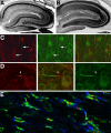
Cellular and subcellular distribution of Kv channels in adult hippocampus. A-C, F: rat, D-E: mouse. A, Double immunofluorescence staining for Kv1.4 (red) and Kv2.1 (green). Note Kv1.4 staining in terminals fields of the medial perforant path in the middle molecular layer of the dentate gyrus, and mossy fiber axons and terminals in s. lucidum of CA3. B, Immunoperoxidase staining for Kv3.1b. C, Double immunofluorescence staining for Kv4.2 (green) and Kv4.3 (red) in dentate gyrus. Note uniform staining for both Kv4 α subunits in granule cell dendrites in molecular layer, and Kv4.3 staining in scattered interneurons. D, Double immunofluorescence staining for Kv7.2 (red) and parvalbumin (green) in CA3. White arrows depict Kv7.2-negative, and red arrow Kv7.2-positive neurons. MF: mossy fibers, sr: s. radiatum, sl: s. lucidum, sp: s. pyramidale, so: s. oriens. Reproduced with permission from reference (59). E, Double immunofluorescence staining for Kv7.2 (green) and DNA (DAPI, blue) in CA1. Reproduced with permission from reference (229). F, In situ hybridization of Kv11.1 (top panel) and Kv11.3 (bottom panel). DG: dentate gyrus. Reproduced with permission from reference (272).
FIG. 3
FIG. 3
Subunit composition and subcellular localization of Nav channel principal and auxiliary subunits in mammalian central neurons. Schematic representation of a single Nav α subunit that forms macromolecular complexes with auxiliary Navβ subunits. Bottom left box, the classification, genetic nomenclature, and subcellular localization of mammalian brain Nav channel principal α subunits. Bottom right box, classification of Navβ auxiliary subunits expressed in mammalian central neurons, and their functional effects on coexpressed Nav α subunits.
FIG. 4
FIG. 4
Cellular and subcellular distribution of Nav channels in mammalian brain and retina. A, B: Immunoperoxidase staining of adult rat hippocampus. Reproduced with permission from reference (100). A, Nav1.1. Note staining in somata and proximal dendrites of dentate granule cells, CA3-CA1 pyramidal cells and interneurons. B, Nav 1.2. Note staining in the mossy fiber pathway within CA3 s. lucidum and in s. radiatum of CA1. C, Double immunofluorescence staining for Nav1 channels (red) and spectrin IVβ (green) in the axonal initial segments (arrows) of pyramidal cells in human temporal neocortex. Reproduced with permission from reference (128). D, Double immunofluorescence staining for Nav1.1 (red) and Nav1.6 (green) in the axonal initial segments of retinal ganglion cells. Arrowhead denotes proximal portion of initial segment positive for Nav1.1 Reproduced with permission from reference (342). E, Triple immunofluorescence staining for nodal Nav1 channels (red), paranodal Caspr (green) and juxtaparanodal Kv1.2 (blue) in adult rat optic nerve. Image courtesy of Dr. Matthew N. Rasband.
FIG. 5
FIG. 5
Subunit composition and subcellular localization of Cav channel principal and auxiliary subunits in mammalian central neurons. Schematic representation of a single Cav α1 subunit that forms macromolecular complexes with auxiliary Cavβ, Cavα2δ, and Cavγ subunits. Bottom left box, the classification, genetic nomenclature, and subcellular localization of mammalian brain Cav channel principal α1 subunits. Bottom right box, classification of Cavβ, Cavα2δ, and Cavγ auxiliary subunits expressed in mammalian central neurons, and their functional effects on coexpressed Nav α subunits.
FIG. 6
FIG. 6
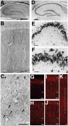
Cellular and subcellular distribution of Cav channels in adult rat brain. A-F: Immunoperoxidase staining. G-K, Immunofluorescence staining. A, D-J: Hippocampus. B, C, K: Neocortex. A, Cav2.1. Note staining in terminals fields of the medial perforant path in the middle molecular layer of the dentate gyrus, and mossy fiber axons and terminals in s. lucidum of CA3. B, Higher magnification view of Cav2.1 staining in CA1. C, Higher magnification view of Cav2.1 staining in CA3, arrows indicate mossy fiber terminals, and arrowheads CA3 pyramidal cell somata. D, Cav1.3. Note staining in somata and proximal dendrites of dentate granule cells, CA3-CA1 pyramidal cells and interneurons. E, Higher magnification view of Cav1.3 staining in CA3-CA2. F, Much higher magnification view of Cav1.3 staining in CA3-CA2. Arrows: base of apical dendrites. Arrowheads: proximal dendrites. G-I: Cav3.1 (G), Cav3.2 (H), and Cav3.3 (I) staining in CA1. J, Cav3.3 staining in subiculum. K, Cav3.3 staining in neocortex. SP, s. pyramidale. SR, s. radiatum. Reproduced with permission from reference: A-C: (359); D-F: (116); G-I: (201).

Similar articles

Cited by

References

    1. Agnew WS, Miller JA, Ellisman MH, Rosenberg RL, Tomiko SA, Levinson SR. The voltage-regulated sodium channel from the electroplax of Electrophorus electricus. Cold Spring Harb Symp Quant Biol. 1983;48(Pt 1):165–179. - PubMed
    1. Ahlijanian MK, Westenbroek RE, Catterall WA. Subunit structure and localization of dihydropyridine-sensitive calcium channels in mammalian brain, spinal cord, and retina. Neuron. 1990;4:819–832. - PubMed
    1. Albensi BC, Ryujin KT, McIntosh JM, Naisbitt SR, Olivera BM, Fillous F. Localization of [125I]omega-conotoxin GVIA binding in human hippocampus and cerebellum. Neuroreport. 1993;4:1331–1334. - PubMed
    1. Albrecht B, Weber K, Pongs O. Characterization of a voltage-activated K-channel gene cluster on human chromosome 12p13. Receptors Channels. 1995;3:213–220. - PubMed
    1. An WF, Bowlby MR, Betty M, Cao J, Ling HP, Mendoza G, Hinson JW, Mattsson KI, Strassle BW, Trimmer JS, Rhodes KJ. Modulation of A-type potassium channels by a family of calcium sensors. Nature. 2000;403:553–556. - PubMed

Publication types

LinkOut - more resources