Review
. 2008 Oct;88(4):1449-90.
doi: 10.1152/physrev.00005.2008.

Ion channels in microbes

Affiliations
Review

Ion channels in microbes

Boris Martinac et al. Physiol Rev. 2008 Oct.

Abstract

Studies of ion channels have for long been dominated by the animalcentric, if not anthropocentric, view of physiology. The structures and activities of ion channels had, however, evolved long before the appearance of complex multicellular organisms on earth. The diversity of ion channels existing in cellular membranes of prokaryotes is a good example. Although at first it may appear as a paradox that most of what we know about the structure of eukaryotic ion channels is based on the structure of bacterial channels, this should not be surprising given the evolutionary relatedness of all living organisms and suitability of microbial cells for structural studies of biological macromolecules in a laboratory environment. Genome sequences of the human as well as various microbial, plant, and animal organisms unambiguously established the evolutionary links, whereas crystallographic studies of the structures of major types of ion channels published over the last decade clearly demonstrated the advantage of using microbes as experimental organisms. The purpose of this review is not only to provide an account of acquired knowledge on microbial ion channels but also to show that the study of microbes and their ion channels may also hold a key to solving unresolved molecular mysteries in the future.


Figures

FIGURE 1
FIGURE 1. Diversity of life forms on Earth
(A) Timeline of the planet Earth showing the early emergence of various life forms on it (reproduced from Woese, 1981 (394) with permission) (B) Universal phylogenetic tree showing the organization of life on Earth in three kingdoms of living organisms: Archaea, Euakarya and Bacteria (modified from Woese, 1994 (393) with permission).
FIGURE 2
FIGURE 2. Different procedures used to generate microbial objects suitable for patch-clamp studies
The starting material (left) – Paramecium, Saccharomyces cerevisiae, Escherichia coli, membrane vesicles and yeast killer toxin – have dimensions on the order of 100, 10, 1, 0.1 and 0.01 µm, respectively. The procedure briefly shown in the center, converts the microbes to objects used successfully for the patch clamp recording. The objects (right) are all 5–15 µm in diameter, except detached cilia from Paramecium, and yeast mitochondria and vacuoles, which are ~2 and ~3 µm in size, respectively. For details of the procedures for preparations of the microbial objects see Saimi et al., 1992 (334) (with permission).
FIGURE 3
FIGURE 3. Bacterial MS channels
(A) The structure of the pentameric MscL channel (left) and a channel monomer (right) from M. tuberculosis according to the 3D structural model (357). (B) A current trace of a single MscL channel reconstituted into azolectin liposomes (w/w protein/lipid of 1:2000) recorded at +30 mV pipette potential. The channel gated more frequently and remained longer open with increase in negative pressure applied to the patch-clamp pipette (trace shown below the channel current trace). (C) 3D structure of the MscS homoheptamer (left) and a channel monomer (right) from E. coli (358). (D) Current traces of two MscS channels reconstituted into azolectin liposomes (w/w protein/lipid of 1:1000) recorded at +30 mV pipette voltage. Increase in pipette suction (trace shown below the channel current trace) caused an increase in the activity of both channels. C and On denote the closed and open state of the n number of channels.
FIGURE 4
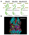
FIGURE 4. MscS-like archaeal channels
(A) MscS monomer (left) and structural models of MscMJ (middle) and MscMJLR (right) monomers based on the MscS crystal structure. Models of the MscMJ and MscMJLR monomers were generated by Swiss-Model (111) and viewed by PyMOL. (B) MscMJLR monomer superimposed and embedded in the 3D structure of MscS of E. coli viewed by PyMOL. α-helices of the MscMJLR monomer are shown in red, β-sheets in yellow, whereas loops are depicted green.
FIGURE 5
FIGURE 5
Topologies of K+-channel subunits. Each channel is a tetramer in forms of a, b, or c, or a dimer of d. (a) A simple form of the subunit comprises two α helices (TM1 and TM2, left and right rods) that traverse the membrane (marked with broken lines, separating the outside above and the cytoplasm below). Between the two, are the pore helix (short rod) and the filter sequence (line between the pore helix and TM2). Certain viral K+ channels, KcsA, Kir, MthK, TvoK, have this topology, though helices preceding TM1 or trailing TM2 exist (not shown) in some of these channels forming structures in the cytoplasm. (b) A common form of subunit comprising 6 transmembrane helices, in which the last two helices and the pore helix (TM5-P-TM6, right portion) form the permeation and filtration core, similar to the structure in a. The preceding four helices (TM1 - TM4, left portion) form a separate domain, housing other structure, such as the voltage sensor. This 6-TM motif is found in KvAP, MVP, KvLm, Kch, MloK, and MmaK. The subunit of the bacterial Na+ channels, NaChBac, also has this structure, although it has a slightly altered filter sequence. (c) The subunit structure of GluR0, a prokaryotic glutamate receptor. The pore helix and the filter sequence are arranged in the direction opposite that of a. The portion outside the membrane forms the glutamate-binding site. (d) The subunit structure of TOK1, the K+ channel of budding yeast. Here the first four TM helices are followed by two core structures, each similar to that in a.
FIGURE 6
FIGURE 6
The crystal structure of KcsA. KcsA is a homotetramer of subunits each with a TM1-P-TM2 core as diagramed in Fig. 5a. a. Viewed from the outside, with each subunit represented in one color and a K+ ion at the center. b. Side view of the membrane-embedded portion of the channel in form of an inverted teepee. Each subunit comprises an outer transmembrane helix (TM1), a short pore helix (P), and an inner transmembrane helix (TM2). Between TM1 and P are residues facing the outside (up), forming a turret. Between P and TM2 is the filter sequence. c, Same view as b, but with the outer TM1 helices removed to show more clearly the inner TM2 helices (red) and pore helices (white). d. The tetramer with the front and back subunit removed to show clearly the turret (white), the filter (GYG, red), and gate region (green). e. Side view of the KcsA tetramer, giving dimension, and showing the two belts of aromatic amino acids facing the membrane (black). From Doyle et al. (1998) (81).

References

    1. Accardi A, Kolmakova-Partensky L, Williams C, Miller C. Ionic currents mediated by a prokaryotic homologue of CLC Cl− channels. J Gen Physiol. 2004;123:109–119. - PMC - PubMed
    1. Ajouz B, Berrier C, Besnard M, Martinac B, Ghazi A. Contributions of the different extramembranous domains of the mechanosensitive ion channel MscL to its response to membrane tension. J Biol Chem. 2000;275:1015–1022. - PubMed
    1. Alabi AA, Bahamonde MI, Jung HJ, Kim JI, Swartz KJ. Portability of paddle motif function and pharmacology in voltage sensors. Nature. 2007;450:370–375. - PMC - PubMed
    1. Alvis SJ, Williamson IM, East JM, Lee AG. Interactions of anionic phospholipids and phosphatidylethanolamine with the potassium channel KcsA. Biophys J. 2003;85:3828–3838. - PMC - PubMed
    1. Anishkin A, Gendel V, Sharifi NA, Chiang CS, Shirinian L, Guy HR, Sukharev S. On the conformation of the COOH-terminal domain of the large mechanosensitive channel MscL. J Gen Physiol. 2003;121:227–244. - PMC - PubMed

Publication types

MeSH terms

LinkOut - more resources