. 2008 Dec 4;456(7222):599-604.
doi: 10.1038/nature07386. Epub 2008 Oct 15.

Centrosome misorientation reduces stem cell division during ageing

Affiliations

Centrosome misorientation reduces stem cell division during ageing

Jun Cheng et al. Nature. .

Abstract

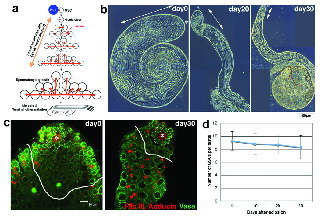
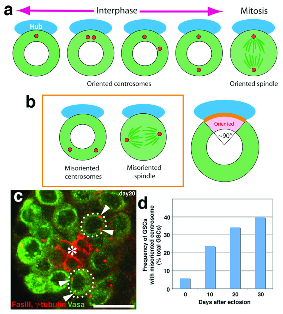
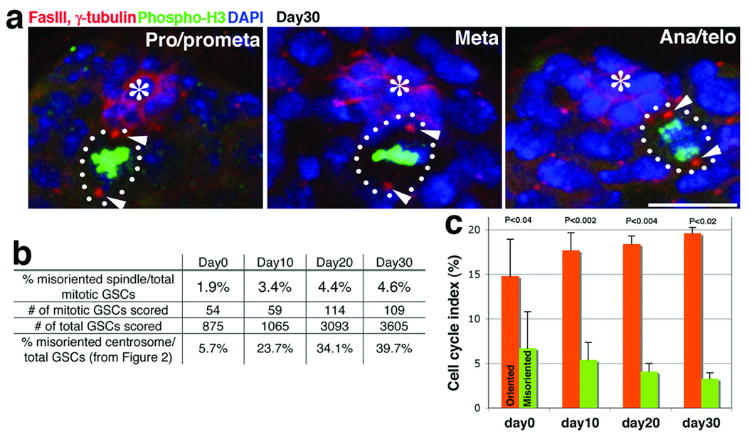
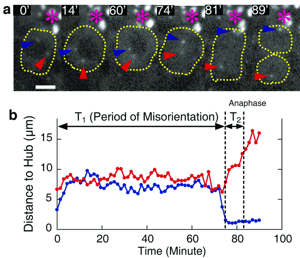
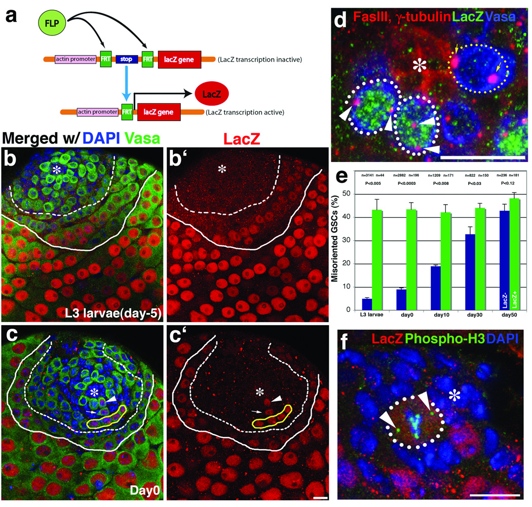
Asymmetric division of adult stem cells generates one self-renewing stem cell and one differentiating cell, thereby maintaining tissue homeostasis. A decline in stem cell function has been proposed to contribute to tissue ageing, although the underlying mechanism is poorly understood. Here we show that changes in the stem cell orientation with respect to the niche during ageing contribute to the decline in spermatogenesis in the male germ line of Drosophila. Throughout the cell cycle, centrosomes in germline stem cells (GSCs) are oriented within their niche and this ensures asymmetric division. We found that GSCs containing misoriented centrosomes accumulate with age and that these GSCs are arrested or delayed in the cell cycle. The cell cycle arrest is transient, and GSCs appear to re-enter the cell cycle on correction of centrosome orientation. On the basis of these findings, we propose that cell cycle arrest associated with centrosome misorientation functions as a mechanism to ensure asymmetric stem cell division, and that the inability of stem cells to maintain correct orientation during ageing contributes to the decline in spermatogenesis. We also show that some of the misoriented GSCs probably originate from dedifferentiation of spermatogonia.


Figures

Figure 1
Figure 1. Drosophila testis undergoes an age-related decline in spermatogenesis
a. Spermatogenesis of Drosophila melanogaster (adopted from Fuller 18). GSCs are supported by the hub cells. Each spermatogonial division is incomplete, and the resultant spermatogonia and spermatocytes are connected by a cytoplasmic bridge, or a ring canal, through which a branched fusome runs. b. Phase microscopy of aging testes. The apical (*) area containing round, relatively early germ cells (arrow) decreases over time. c. Number of GSCs (surrounded by dotted line) remains constant with age. White lines separate spermatogonia and spermatocytes. Red, Fasciclin III (Fas III; hub) and Adducin (fusome); Green, Vasa (germ cells). Hub (*). d. The number of GSCs (± SD). n > 50 testes were counted for each time point.
Figure 2
Figure 2. Misoriented GSCs increase with age
a. Schematic diagram of centrosome positioning during the cell cycle. b. Left panel; the definition of misoriented centrosomes and spindle. Right panel; scoring criterion. Centrosomes were scored to be oriented when one of two centrosomes is in the pink area close to the hub-GSC junction (orange line). c. An example of testis (20-day old) containing GSCs with misoriented centrosomes. Red, Fas III and γ-tubulin (centrosome); Green, Vasa. Hub(*). Bar, 10 µm. d. Frequency of GSCs with misoriented centrosomes increases with age (n > 275 GSCs for each time point). The same trend was observed in more than three separate experiments, including conditions with different culture media and temperature (22°C–25°C).
Figure 3
Figure 3. Misoriented GSCs divide less frequently compared to oriented GSCs
a. Spindles remain oriented throughout mitosis even at day 30. Red, Fas III and γ-tubulin; Green, Thr3-phosphorylated histone H3 (phospho-H3; mitotic chromatin); Blue, DAPI. Hub(*). Bar, 10 µm. b. Frequency of misoriented spindle remained low for up to 30 days of age. The percentage of misoriented interphase cells was taken from Figure 2C for comparison. c. Pulse-labeling of centrosomes by heatshock-induced GFP-PACT expression confirmed the low cell cycle activity of misoriented GSCs. The percentage of GSCs committed to the G1/S transition during the labeling period is shown (mean ± SD; n > 110 labeled GSCs for each point, equivalent to n > 750 total GSCs per data point). P values of t-test (two-tail) are shown.
Figure 4
Figure 4. Misoriented GSCs correct centrosome orientation prior to mitosis
a. Frames from time-lapse imaging of GSC cell division with misoriented centrosome (Movie is available on line, Supplemental Movie 1). Centrosome movement in a GSC was tracked by mCherry-Sas6. Numbers represent minutes from the start point of recording. Re-orientation occurred around 74 min, and anaphase around 81 min. Hub (*), and two centrosomes (red and blue arrowheads). b. Tracking of the centrosome movement from the same time-lapse imaging. Red and blue lines represent the movement of centrosomes with red and blue arrowheads shown in (a), respectively.
Figure 5
Figure 5. Dedifferentiated GSCs have higher frequency of centrosome misorientation
a. Strategy to label germ cells that committed to differentiation program through FLP-based recombination. b and c. LacZ staining of testes from an L3 larva (b) and a day 0 adult (c). Week LacZ expression starts around 4–8 cell spermatogonia (dashed line), and become stronger in spermatocytes (solid line). Arrowhead in (c) shows LacZ+ GSC, with its progeny (gonialblast shown by arrow and 4 cell spermatogonia surrounded by yellow line). Red, LacZ; Green, Vasa; Blue, DAPI. Hub (*). Bar, 10 µm. d. An example of testis with two misoriented LacZ+ GSCs (white dotted line, with arrowheads indicating misoriented centrosomes) and a correctly oriented LacZ GSC (yellow dotted line, with arrows indicating properly oriented centrosomes). Red, Fas III and γ-tubulin; Green, LacZ; Blue, Vasa. e. LacZ+ GSCs have a high frequency of misorientation (mean ± SD). P values of t-test (two-tail) are shown. f. LacZ+ GSCs are correctly oriented during mitosis. 4th chromosomes that segregate before anaphase mark spindle poles (arrowheads). Red, LacZ; Green, phospho-H3; Blue, DAPI.
Figure 6
Figure 6. Evidence that LacZ+ GSCs are generated by dedifferentiation
a. Summary of LacZ+ GSC frequencies with or without X-ray irradiation. Flies were irradiated at day 0 of age, and analyzed at day 5. The actual and expected frequencies of singlet, doublet, triplet, and quadruplet (or more) are shown. N; number of GSCs scored. The expected frequencies were calculated as described in Full Methods. b. An example of testis apical tip (irradiated with 2000 rad) containing a triplet (arrowheads: 3 LacZ+ GSCs are locating next each other). Red; LacZ, Green; Vasa, Blue; DAPI. Bar; 10µm. Hub(*). c. and d. Examples of LacZ+ (c) and LacZ− (d) dedifferentiating spermatogonia (surrounded by dotted lines), with multiple ring canals (arrowheads) and disintegrating fusomes (arrows), observed 24 hours after irradiation. Red, LacZ and adducin; Green, Anillin (ring canal and nucleus); Blue, Vasa. Bar; 10µm.

Comment in

References

    1. Morrison SJ, Kimble J. Nature. 2006;441(7097):1068. - PubMed
    1. Spradling A, Drummond-Barbosa D, Kai T. Nature. 2001;414(6859):98. - PubMed
    2. Fuchs E, Tumbar T, Guasch G. Cell. 2004;116(6):769. - PubMed
    1. Yamashita YM, Fuller MT, Jones DL. J Cell Sci. 2005;118(Pt 4):665. - PubMed
    1. Rando TA. Nature. 2006;441(7097):1080. - PubMed
    1. Serrano M, Blasco MA. Nat Rev Mol Cell Biol. 2007;8(9):715. - PubMed
    2. Sharpless NE, Depinho RA. Nat Rev Mol Cell Biol. 2007;8(9):703. - PubMed

Publication types