. 2008 Nov 19;27(22):2943-54.
doi: 10.1038/emboj.2008.211. Epub 2008 Oct 16.

Clustering of VASP actively drives processive, WH2 domain-mediated actin filament elongation

Affiliations

Clustering of VASP actively drives processive, WH2 domain-mediated actin filament elongation

Dennis Breitsprecher et al. EMBO J. .

Abstract

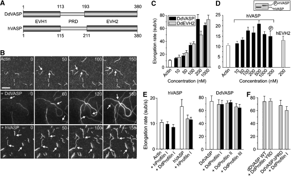
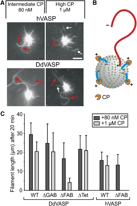
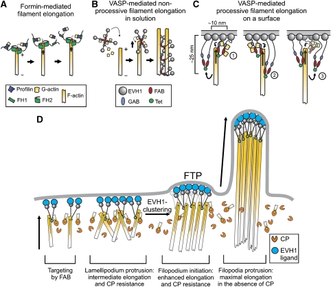
Vasodilator-stimulated phosphoprotein (VASP) is a key regulator of dynamic actin structures like filopodia and lamellipodia, but its precise function in their formation is controversial. Using in vitro TIRF microscopy, we show for the first time that both human and Dictyostelium VASP are directly involved in accelerating filament elongation by delivering monomeric actin to the growing barbed end. In solution, DdVASP markedly accelerated actin filament elongation in a concentration-dependent manner but was inhibited by low concentrations of capping protein (CP). In striking contrast, VASP clustered on functionalized beads switched to processive filament elongation that became insensitive even to very high concentrations of CP. Supplemented with the in vivo analysis of VASP mutants and an EM structure of the protein, we propose a mechanism by which membrane-associated VASP oligomers use their WH2 domains to effect both the tethering of actin filaments and their processive elongation in sites of active actin assembly.


Figures

Figure 1
Figure 1
Analysis of VASP-mediated actin assembly by TIRF microscopy. (A) D. discoideum DdVASP and human hVASP share a similar domain organization encompassing an N-terminal EVH1, a central proline-rich region (PRD) and C-terminal EVH2 domain. Numbers indicate amino-acid residues. (B) VASP accelerates actin assembly in vitro. A concentration of 1 μM ATP-actin and 0.3 μM Alexa-Fluor-488 labelled ATP-actin (from now on referred to as 488 actin) was polymerized in the presence or absence of either 200 nM DdVASP or 200 nM hVASP on NEM-myosin II-coated glass slides in 1 × TIRF buffer containing 10 mM imidazole, 50 mM KCl, 1 mM MgCl2, 1 mM EGTA, 0.2 mM ATP, 50 mM DTT, 15 mM glucose, 20 μg/ml catalase, 100 μg/ml glucose oxidase and 0.5% methylcellulose (4000 cP), pH 7.4. Time-lapse micrographs of the assembly reaction are shown. The time is indicated in seconds on top. Arrows mark barbed ends during filament elongation. Scale bar, 10 μm. (C) Concentration dependence of the elongation rates of DdVASP-mediated actin assembly by the full-length protein and the EVH2 domain (DdEVH2). (D) Comparison of the actin filament elongation rates in the presence of untreated or PKA phosphorylated WT hVASP and the hEVH2 domain alone. Inset shows SDS–PAGE of Coomassie Blue-stained non-phosphorylated and PKA-phosphorylated hVASP. (E) Profilin does not accelerate VASP-mediated actin assembly. The elongation rates of actin and either 200 nM hVASP (left) or 200 nM DdVASP (right) in the presence of 2 μM (grey bars) or 5 μM (black bars) of the indicated profilin isoforms are shown. (F) Profilin–PRD interaction is not required for VASP-mediated filament elongation. The elongation rate of 200 nM DdVASP was not affected by addition of 5 μM profilin Y6D mutant. Data in C–F correspond to means±s.e.m.
Figure 2
Figure 2
Analysis of DdVASP mutants lacking functional domains. (A) Localization of the GAB, FAB and Tet motifs within the EVH2 domain of DdVASP. Numbers indicate deleted amino-acid residues in the mutants. Mutant DdVASPΔGAB/FAB lacks residues 198–220 and 262–284. (B) Both WH2 domain-like actin-binding motifs can contribute to actin assembly. Time-lapse micrographs of the assembly of 1 μM ATP-actin and 0.3 μM 488 actin in 1 × TIRF buffer in the presence of 200 nM DdVASPΔGAB (blue arrows), DdVASPΔFAB (red arrows) and DdVASPΔTet (green arrows). Time and scale as in Figure 1B. (C) Comparison of the actin filament elongation rates in the presence of different DdVASP mutants at the concentrations indicated. The assembly rates for untagged or GST-tagged DdVASPΔTet constructs were virtually identical. Data correspond to means±s.e.m. (DG) The FAB motif and tetramerization of VASP are required for robust filament side binding. A concentration of 2 μM actin was polymerized in 1 × TIRF buffer lacking methylcellulose on glass slides coated with NEM-myosin II in the presence of 1 μM of the GST-tagged DdVASP WT, ΔGAB, ΔFAB constructs and of untagged monomeric ΔTet mutant. After 20 min, the samples were fixed and stained with anti-GST or DdVASP polyclonal antibodies. Primary antibodies were visualized with Alexa-Fluor-488-conjugated goat-anti-rabbit antibodies and F-actin was stained with rhodamine-phalloidin. Confocal images of the specimens are shown. As DdVASPΔFAB and DdVASPΔTet mutants lacked bundling activity, filament bundling was induced by 10 μM poly-L-lysine (F, G).
Figure 3
Figure 3
Capping protein inhibits VASP-mediated actin assembly in solution. (A, D) TIRF images of actin filaments polymerized for 10 min with either DdVASP or hVASP (200 nM each) in the presence and absence of Cap32/34 or CapZ (40 nM each) in 1 × TIRF buffer, respectively. White arrows in the TIRF images (left) and corresponding kymographs (right) indicate filaments that stopped growing in the presence of CP. The growth of non-capped filaments is shown for comparison. Scale bar, 10 μm. (B, E) Cap32/34 and CapZ decrease the average length of actin filaments formed in the presence of DdVASP or hVASP. Filaments formed in the presence of VASP were longer than the actin control. (C, F) The time to growth arrest is virtually independent of VASP. The times of filament elongation until capping were calculated from the elongation rates of VASP-mediated actin assembly. The lengths of at least 150 filaments for each condition were measured. Note different scales in B and E. Data correspond to means±s.e.m.
Figure 4
Figure 4
Clustering of VASP promotes processive actin filament elongation. (A) Actin assembly of 1 μM actin and 0.3 μM 488 actin in 1 × TIRF buffer on 2 μm polystyrene beads coated with the DdVASP constructs indicated. Blue arrows indicate filaments that grew with their barbed ends pointing away from the beads and red arrows highlight fast growing, buckling filaments with their barbed ends attached to the bead surface. Determination of the elongation rates (right) revealed two different filament populations. The blue and red lines correspond to the filaments marked in the time-lapse micrographs. The black line is shown as a control and corresponds to the elongation rate of a single actin filament that grew spontaneously in solution beside the beads. Time is shown in seconds. Scale bar, 10 μm. (B) Proposed mechanism leading to different filament populations on VASP-coated beads. + indicates a barbed and − indicates a pointed end. (C) Mutants DdVASPΔGAB, −ΔFAB and −ΔTet produced buckling filaments that grew with fast elongation rates, whereas DdVASPΔGAB/FAB failed to recruit actin and to assemble filaments. Scale bar and arrows are as in (A). (D) Quantification of the fraction of processively growing filaments on VASP-coated beads. For each box, at least 30 beads from six independent experiments were analysed. Data correspond to means per bead±s.e.m. (E) Analysis of DdVASP density on functionalized polystyrene beads. Average separation distance of DdVASP tetramers on functionalized beads depends on the amount of DdVASP used for coating (left). The dashed line indicates the maximal distance at which robust processive filament elongation was observed. At the minimum concentration required for processive filament elongation, more than 50% of the beads produce buckling filaments (right). For each data point, at least 150 beads were analysed. (F) Electron microscopic analysis of the VASP tetramer. High magnification galleries of recombinant mVASP (left) and high magnification galleries of an mVASP deletion mutant lacking amino-acid residues 118–304 but still containing the tetramerization domain (right) are shown. Scale bars, 50 nm. (G) Distributions of molecular lengths of WT and mutant mVASP proteins determined from electron micrographs such as those displayed in the shown galleries in (F). The distance between the two furthermost globular domains was measured. n=124 for mVASP and n=116 for mutant mVASPΔ118–304.
Figure 5
Figure 5
Clustered VASP promotes processive filament elongation in the presence of CP. (A) Both human- and DdVASP-coated beads assemble long actin filaments in the presence of up to 1 μM of species-specific CP. Red arrows indicate growing filaments, small white arrows highlight short capped filaments that detached from the beads. Scale bar, 5 μm. (B) Scheme illustrating the elimination of filaments growing with their barbed ends pointing away from the bead by CP. Only filaments assembled by VASP at the surface remain. (C) The FAB motifs of DdVASP and hVASP are critical for CP resistance upon clustering. For each mutant, the 20% longest filaments growing on coated beads were analysed after 20 min. Mutant hVASPΔFAB failed to produce filaments in the presence of 1 μM CP. For each box, at least 20 filaments were analysed. Data correspond to means±s.e.m.
Figure 6
Figure 6
Ability of VASP constructs to restore filopodia formation and cell migration in Dictyostelium VASP-null cells. (A) 3D reconstitutions of phalloidin-labelled cells indicated are shown. Rescue of filopodia formation was not observed for the VASPΔFAB construct fused to GFP. Typical Ax2-WT and VASP-null cells are shown as controls. Scale bar, 10 μm. (B) FAB is critical for VASP targeting to the tip of the leading edge. Confocal sections show accumulation of the indicated GFP-tagged VASP constructs during protrusion of the leading edge, white arrows indicate direction of protrusion during random motility (left panel). Insets show localization of GFP-tagged constructs in filopodia. Intensity profiles corresponding to a linear 8-bit grey scale of the GFP fluorescence in boxed regions are shown (right panel). Scale bar, 10 μm. (C) Cell motility of WT, VASP-null and reconstituted cells. Quantification data represent results from three independent experiments. For each cell line, at least 25 cells were analysed. Boxes indicate 25th percentile, median and 75th percentile of all values; error bars indicate 10th and 90th percentile.
Figure 7
Figure 7
Models of VASP-mediated actin assembly. (A) Processive filament elongation by formins is shown for comparison. (B) Proposed mechanism for non-processive filament elongation by VASP in solution. VASP tetramers loaded with actin monomers hit a free barbed end, transiently bind and deliver bound actin subunits to it, resulting in non-processive filament elongation. Subsequent side binding of VASP results in decoration of the filament and mediates bundle formation. (C) Proposed mechanism for processive filament elongation on a surface: (1) VASP tetramers tethered to the surface bind to actin and deliver monomers through their WH2 domains to the barbed end. (2) After delivery, VASP remains bound to the side of the filament as the barbed end elongates in response to the delivery of actin monomers by other VASP molecules. Owing to its flexible architecture, VASP may stay attached to the side of the filament up to 20–30 nm away from the surface. (3) Stretched VASP molecules eventually detach from the filament due to continuous elongation of the barbed end and are afterwards available for a new cycle of actin addition. During the detachment period, the growing filament is constantly tethered to the surface by other VASP molecules. Decoration of the barbed end region by multiple VASP molecules is presumed to prevent access of CP to the filament barbed end. As filament-growth-induced detachment of VASP does not occur in solution, processive elongation can be accomplished only on a surface. (D) Functions of VASP in cell protrusions. VASP molecules docked on the membrane through their EVH1 domains anchor actin filaments through their WH2 domains. This anchorage may in turn stabilize the localization of VASP to the membrane. Clustering of VASP allows lamellipodium protrusion in the presence of CP, which itself might serve to eliminate unproductive filaments. Stronger clustering mediated by an EVH1 ligand may trigger the formation of the filopodium tip complex (FTP), allowing the transition from lamellipodium to filopodium formation. For the sake of simplicity, VASP is illustrated only as a dimer in C and D.

References

    1. Aebi U, Baschong W (2006) Glycerol spraying/low-angle rotary metal shadowing. In Cell Biology—A Laboratory Handbook, J Celis (ed), 3rd ed, pp 241–246. London: Elsevier
    1. Applewhite DA, Barzik M, Kojima S, Svitkina TM, Gertler FB, Borisy GG (2007) Ena/VASP proteins have an anti-capping independent function in filopodia formation. Mol Biol Cell 18: 2579–2591 - PMC - PubMed
    1. Bachmann C, Fischer L, Walter U, Reinhard M (1999) The EVH2 domain of the vasodilator-stimulated phosphoprotein mediates tetramerization, F-actin binding, and actin bundle formation. J Biol Chem 274: 23549–23557 - PubMed
    1. Ball LJ, Kuhne R, Hoffmann B, Hafner A, Schmieder P, Volkmer-Engert R, Hof M, Wahl M, Schneider-Mergener J, Walter U, Oschkinat H, Jarchau T (2000) Dual epitope recognition by the VASP EVH1 domain modulates polyproline ligand specificity and binding affinity. EMBO J 19: 4903–4914 - PMC - PubMed
    1. Barzik M, Kotova TI, Higgs HN, Hazelwood L, Hanein D, Gertler FB, Schafer DA (2005) Ena/VASP proteins enhance actin polymerization in the presence of barbed end capping proteins. J Biol Chem 280: 28653–28662 - PMC - PubMed

Publication types

MeSH terms