Review
. 2008 Nov;8(11):880-7.
doi: 10.1038/nrc2505. Epub 2008 Oct 16.

VEGF-A splicing: the key to anti-angiogenic therapeutics?

Affiliations
Review

VEGF-A splicing: the key to anti-angiogenic therapeutics?

Steven J Harper et al. Nat Rev Cancer. 2008 Nov.

Abstract

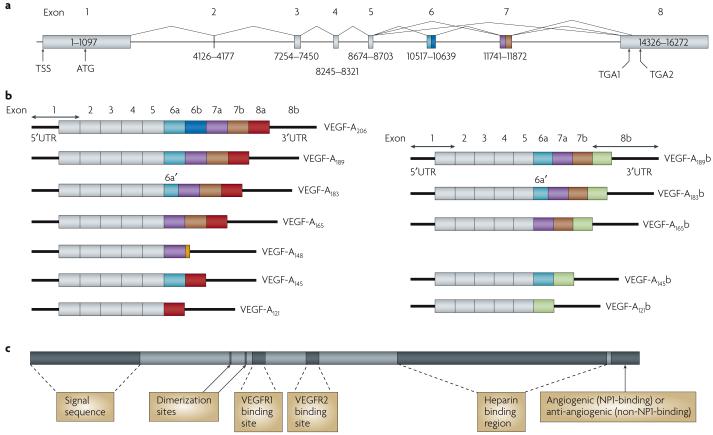
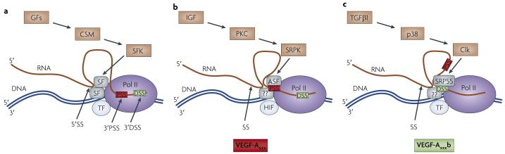
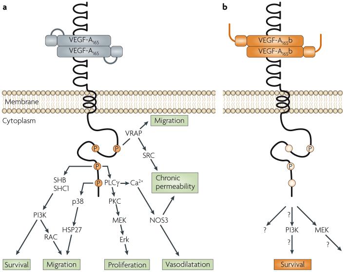
The physiology of microvessels limits the growth and development of tumours. Tumours gain nutrients and excrete waste through growth-associated microvessels. New anticancer therapies target this microvasculature by inhibiting vascular endothelial growth factor A (VEGF-A) splice isoforms that promote microvessel growth. However, certain VEGF-A splice isoforms in normal tissues inhibit growth of microvessels. Thus, it is the VEGF-A isoform balance, which is controlled by mRNA splicing, that orchestrates angiogenesis. Here, we highlight the functional differences between the pro-angiogenic and the anti-angiogenic VEGF-A isoform families and the potential to harness the synthetic capacity of cancer cells to produce factors that inhibit, rather than aid, cancer growth.


Figures

Figure 1
Figure 1. Protein and mRNA products of human vascular endothelial growth factor A (VEGF-A)
a | Gene structure of human VEGF-A. VEGF-A spans 16,272 bp of chromosome 6p12 and consists of eight exons. Alternate 5′ and 3′ splice site selection in exons 6, 7 and 8 generate multiple isoforms. Exons 6 and 7 encode heparin binding domains. The transcriptional start site (TSS) and translational start site (ATG) in exon 1 are indicated. Alternative stop codons within exon 8 are also indicated (TGA1 and TGA2). b | Alternative splicing can occur either at the 5′ donor splice site (for example, VEGF-A189 versus VEGF-A206) or the 3′ acceptor splice site (for example, VEGF-A189 versus VEGF-A165). Two mRNA isoform families are generated. The pro-angiogenic isoforms (VEGF-Axxx, left) are generated by proximal splice site (PSS) selection in exon 8 and the anti-angiogenic family (VEGF-Axxxb, right) from exon 8 distal splice site (DSS) choice. Thus, VEGF-A165, formed by PSS selection in exon 8, has VEGF-A165b as its DSS sister isoform, the DSS-selected mRNA encoding a protein of exactly the same length. Exon 6a’ occurs in VEGF-A183 as a result of a conserved alternative splicing donor site in exon 6a and is 18 bp shorter than full-length exon 6a. VEGF-A148 is a truncated isoform splicing from exon 7a into exon 8a out of frame and resulting in a premature stop codon. VEGF-A206b has not yet been identified. c | Protein structure of VEGF-A containing the dimerization sites and binding sites for heparin, VEGF-A receptor 1 (VEGFR1; encoded by exon 3) and VEGFR2 (encoded by exon 4), which are present in all isoforms. The six amino acids at the extreme carboxyl terminus of the protein can be either pro-angiogenic (CDKPRR, encoded by exon 8a) or anti-angiogenic (SLTRKD, encoded by exon 8b). The epitopes recognized by most commercial antibodies are in the region of the VEGF-A receptor-binding domains, present in VEGF-A isoforms of both families. UTR, untranslated region.
Figure 2
Figure 2. Vascular endothelial growth factor A (VEGF-A) C’ terminal splicing regulation
a | The C’ terminal domain of RNA polymerase II (Pol II) interacts with both transcription factors (TFs) and splicing factors (SFs). SFs are recruited to the transcriptional machinery owing to their interaction with Pol II,. These SFs recognize cis-acting RNA splicing sequences in the pre-mRNA and both splicing sites (SS) — 5′ donor (5′SS) or 3′ acceptor sites — can be recognized. Both 3′ proximal SS (3′PSS) and distal SS (3′DSS) are indicated. The particular splicing factors recruited are dependent on the sequence. These SFs can be regulated by SF kinases (SFKs), which are regulated by cell signalling molecules (CSMs) downstream of growth factors (GFs). b | Regulation of VEGF-A C’ terminal PSS selection by insulin-like growth factor (IGF). IGF activates protein kinase C (PKC), which results in phosphorylation of SR protein kinases (SRPKs). These can activate the ASF-SF2 splicing factor, which favours PSS selection. This process may be dependent on the presence of hypoxia-inducible factor (HIF), a transcription factor involved in VEGF-Axxx upregulation. Other SFs and kinases may also be involved in PSS and DSS selection, denoted by ‘??’. c | Factors affecting VEGF-A C’ terminal DSS selection. Transforming growth factor β1 (TGFβ1) results in p38 mitogen-activated protein kinase activation and subsequent activation of the kinases CLK1 and CLK4. CLK1 and CLK4 phosphorylate the splicing factor SRP55, resulting in DSS selection and production of VEGF-Axxxb​. It is also possible that ASF-SF2 is inactivated by CLK1 and CLK4, or that phosphorylation of the SFs could change their location, degradation or binding affinity. This scheme summarizes the limited data available.
Figure 3
Figure 3. Signalling pathways downstream of vascular endothelial growth factor (VEGF-A)xxx and VEGF-Axxxb
a | The VEGF-Axxx-mediated angiogenic response acts primarily through VEGF receptor 2 (VEGFR2) to initiate multiple downstream pathways,. b | VEGF-A165b results in transient, weak phosphorylation and the downstream signalling (denoted ‘?’) from such qualitatively different phosphorylation is largely unknown (see REF. for details). Erk, extracellular signal-regulated kinase; HSP27, heat shock protein 27; NOS3, endothelial nitric oxide synthase; PI3K, phosphoinositide 3-kinase; PKC, protein kinase C; RAC, Ras-related C3 botulinum toxin substrate; PLCγ, phospholipase Cγ; SHB, SH2 domain-containing adaptor protein B; SHC1, SH2 domain-containing transforming protein 1; VRAP, VEGF receptor-associated protein.
Figure 4
Figure 4. The structure of vascular endothelial growth factor A (VEGF-A)
a | Crystal structure of amino acids 4-108 of VEGF-A, which are present in all isoforms. The crystal structure of the full-length VEGF-A protein is not known as a hinge region after amino acid 108 prevents crystallization. Modified, with permission, from REF. © 1999 Elsevier B.V. b | Amino acids 4-108 of VEGF-A are shown along with the crystal structure of the final 55 residues. Crystallization of the final 55 residues of VEGF-A165 indicates two cysteine (C)-bonded double anti-parallel β sheet structures (brown arrows) separated by an α helix (blue cylinder). This structure is highly mobile and rotates around the hinge, and could pass through the VEGF receptor 2 (VEGFR2) binding region but not the VEGFR1 region (yellow circles),. c | Proposed structure of amino acids 108-165 of VEGF-A165. The C’ terminal six residues include a cysteine with two positively charged arginines (RR) that are proposed to interact with the VEGFR binding domain to activate intracellular torsional rotation of VEGFR2. The RR motif therefore acts as a molecular switch by inducing a conformational change in VEGFR2. A disulphide bond (shown in orange) between cysteines 146 and 160 is required for VEGF-A165 activity and ensures that the C terminus is maintained at close proximity to the neuropilin 1 binding domain (NPBD). d | Proposed structure of amino acids 108-165 of VEGF-A165b. The C’ terminal cysteine and the positively charged RR motif present in VEGF-A165 are replaced by a serine (S) and a neutral DK motif on VEGF-A165b respectively. Although the VEGFR binding domain is present, the cysteine disulphide bond is absent. Thus, the molecular interaction with VEGFR is likely to be significantly different.
Figure 5
Figure 5. Vascular endothelial growth factor A (VEGF-A)165b and VEGF-A165 interaction with VEGF receptor 2 (VEGFR2)
a | The VEGFR2 binding site of VEGF-A165 interacts with the VEGFR2 extracellular domain. VEGF-A165 functions as a dimer and promotes the formation of VEGFR2 dimers (only one receptor is shown here for clarity) resulting in activation of the split kinase domains (green lines) and the phosphorylation of tyrosine residues 951, 1152 and 1214 (orange) and 1054 (purple). The charged residues at the carboxy-terminal end of the VEGF-A165 molecule (omitted for clarity) are required for VEGFR activation and, in receptor tyrosine kinases, this is thought to occur through torsional rotation of the intracellular domain bringing together the split kinase domains. Tyrosine 1054 is located at the mouth of the ATP binding pocket of the tyrosine kinase and, once phosphorylated, prevents the binding pocket from closing, thus resulting in a stable open structure. This results in formation of a persistently functional kinase from the split kinase domains, resulting in sustained cis- and trans- phosphorylation of the tyrosine residues on the intracellular tail, even in the presence of phosphatases. Robust tyrosine phosphorylation also results in the activation of angiogenic signalling pathways (FIG. 3). b | VEGF-A165b binds the VEGFR2 binding site with equal affinity to VEGF-A165 but does not bind neuropilin 1 (NRP1). The C’ terminus of VEGF-A165b is neutral and there is insufficient torsional rotation for tyrosine 1054 to be phosphorylated, although weak phosphorylation of the other tyrosines can occur. Thus, the ATP binding pocket closes and the phosphorylated tyrosines can be rapidly dephosphorylated by phosphatases and trafficked much more quickly. As a result, angiogenic signalling pathways are not activated.

References

    1. Mendel DB, et al. In vivo antitumor activity of SU11248, a novel tyrosine kinase inhibitor targeting vascular endothelial growth factor and platelet-derived growth factor receptors: determination of a pharmacokinetic/pharmacodynamic relationship. Clin. Cancer Res. 2003;9:327–337. - PubMed
    1. Motzer RJ, et al. Sunitinib versus interferon α in metastatic renal-cell carcinoma. N. Engl. J. Med. 2007;356:115–124. - PubMed
    1. Hurwitz H, et al. Bevacizumab plus irinotecan, fluorouracil, and leucovorin for metastatic colorectal cancer. N. Engl. J. Med. 2004;350:2335–2342. - PubMed
    1. Ferrara N. Role of vascular endothelial growth factor in physiologic and pathologic angiogenesis: therapeutic implications. Semin. Oncol. 2002;29:10–14. - PubMed
    1. Ferrara N, Henzel WJ. Pituitary follicular cells secrete a novel heparin-binding growth factor specific for vascular endothelial cells. Biochem. Biophys. Res. Comm. 1989;161:851–858. - PubMed

MeSH terms

Substances