. 2008 Oct 16;455(7215):984-7.
doi: 10.1038/nature07439.

A role for the two-helix finger of the SecA ATPase in protein translocation

Affiliations

A role for the two-helix finger of the SecA ATPase in protein translocation

Karl J Erlandson et al. Nature. .

Abstract

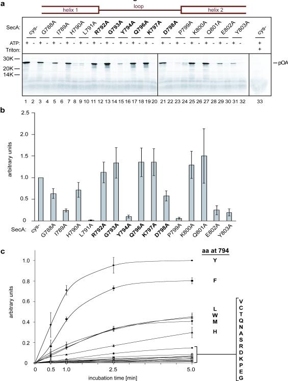
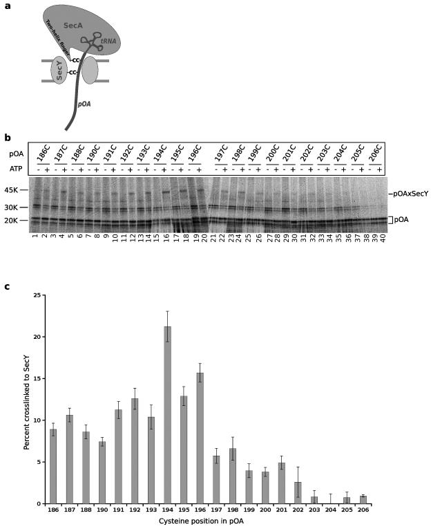
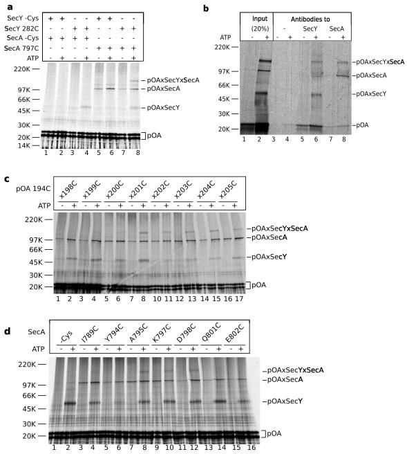
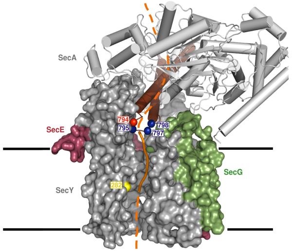
An important step in the biosynthesis of many proteins is their partial or complete translocation across the plasma membrane in prokaryotes or the endoplasmic reticulum membrane in eukaryotes. In bacteria, secretory proteins are generally translocated after completion of their synthesis by the interaction of the cytoplasmic ATPase SecA and a protein-conducting channel formed by the SecY complex. How SecA moves substrates through the SecY channel is unclear. However, a recent structure of a SecA-SecY complex raises the possibility that the polypeptide chain is moved by a two-helix finger domain of SecA that is inserted into the cytoplasmic opening of the SecY channel. Here we have used disulphide-bridge crosslinking to show that the loop at the tip of the two-helix finger of Escherichia coli SecA interacts with a polypeptide chain right at the entrance into the SecY pore. Mutagenesis demonstrates that a tyrosine in the loop is particularly important for translocation, but can be replaced by some other bulky, hydrophobic residues. We propose that the two-helix finger of SecA moves a polypeptide chain into the SecY channel with the tyrosine providing the major contact with the substrate, a mechanism analogous to that suggested for hexameric, protein-translocating ATPases.


Figures

Figure 1
Figure 1. An essential tyrosine at the tip of the two-helix finger
a, Residues in SecA’s two-helix finger were individually mutated to alanines. Position 795 is an alanine in the wild type and cysteine-lacking (Cys-) proteins and therefore remained unchanged. Residues in the helices and loops are indicated in the scheme on top and loop residues are also highlighted in bold. The mutants were purified and tested for translocation activity by incubation for 5 min at 37°C in the presence or absence of ATP with in vitro synthesized 35S-labeled pOA and proteoliposomes containing purified SecY complex. After treatment with proteinase K, the samples were separated by SDS-PAGE and analyzed by autoradiography. The Cys- mutant served as positive control. For each mutant, samples were also treated with protease in the presence of Triton X-100 to disrupt the membrane (shown here in lane 33 for the Cys- mutant). b, Quantification of three experiments performed as in a (mean and standard errors). The data were normalized with respect to the Cys- mutant. c, Translocation kinetics of SecA mutants in which Tyr794 was replaced with other residues (in one-letter code). Shown are the mean and standard deviation of three experiments, normalized to the 5-min data point of the Cys- mutant, which has the wild type tyrosine (Y) in the loop.
Figure 2
Figure 2. Contact of a translocation intermediate with the pore of SecY
a, Scheme of the crosslinking strategy. A tRNA-associated fragment of 35S-proOmpA (pOA) was synthesized in vitro and translocated by SecA into the SecY channel. The bulky tRNA prevents complete translocation. Two cysteines (C) were introduced into pOA, one for crosslinking to a cysteine in the two-helix finger of SecA and one for crosslinking to a cysteine in SecY. b, Translocation substrates (pOA:tRNA) of 206 residues containing single cysteines at the indicated positions were incubated in the absence or presence of ATP with a cysteine-free SecA and proteoliposomes containing purified SecY complex. SecY carried a single cysteine in the pore ring at position 282. After oxidation with Cu2+-phenanthroline, the samples were treated with NEM and RNase A, and analyzed by non-reducing SDS-PAGE and autoradiography. The positions of free and crosslinked pOA (pOA and pOAxSecY) are indicated. c, Quantification of three experiments performed as in b (mean and standard deviation).
Figure 3
Figure 3. The two-helix finger of SecA interacts with a translocating substrate
a, Translocation substrates (pOA:tRNA) of 206 residues containing cysteines at positions 194 and 201 were incubated with SecA and proteoliposomes containing SecY complex in the absence or presence of ATP. The SecA proteins either lacked a cysteine or contained a cysteine at the tip of the two-helix finger (position 797). SecY lacked a cysteine or contained a cysteine in the pore ring (position 282). After oxidation with Cu2+-phenanthroline, the samples were treated with NEM and RNase A, and analyzed by non-reducing SDS-PAGE and autoradiography. The positions of free and crosslinked pOA (pOA, pOAxSecY, pOAxSecA, and pOAxSecYxSecA) are indicated. b, As in a, but the proteoliposomes were sedimented before addition of the oxidant. SecA contained a cysteine at position 797. After solubilization in DDM, 20% of each sample was analyzed directly (lanes 1 and 2), while the remainder was incubated with protein G beads either without antibody or with SecA- or SecY- antibodies (lanes 3-8). c, As in a, but the proteoliposomes containing SecY with a cysteine at position 282 were incubated with a SecA mutant containing a single cysteine at position 797 and pOA:tRNA with a cysteine at position 194 and a second cysteine at the indicated position. The samples were sedimented before addition of the oxidant. d, As in c, but the proteoliposomes containing SecY with a cysteine at position 282 were incubated with pOA:tRNA containing cysteines at positions 194 and 201 and SecA mutants containing single cysteines at the indicated positions.
Figure 4
Figure 4. Model of SecA translocating a polypeptide into the SecY channel
A segment of a translocation substrate was modeled in orange in the T. maritima SecA-SecY complex structure (TM2b and residues 72-76 were removed for clarity) . The segment from the tip of the two-helix finger of SecA to the pore ring of SecY is shown as a solid line, the other segments as broken lines. Translocating proOmpA containing two cysteines could be double-crosslinked to position 282 of SecY (yellow) and to SecA positions in blue at the tip of the two-helix finger (brown). The essential tyrosine in the loop between the two helices is shown in red. SecY is shown as a space-filling model and SecA as a cartoon. The numbers correspond to the positions in E.coli SecA and SecY. The membrane boundaries are indicated.

Comment in

References

    1. Rapoport TA. Protein translocation across the eukaryotic endoplasmic reticulum and bacterial plasma membranes. Nature. 2007;450:663–669. - PubMed
    1. Brundage L, Hendrick JP, Schiebel E, Driessen AJM, Wickner W. The purified E. coli integral membrane protein SecY/E Is sufficient for reconstitution of SecA-dependent precursor proteintranslocation. Cell. 1990;62:649–657. - PubMed
    1. Zimmer J, Nam Y, Rapoport TA. Crystal structure of the protein-translocation complex formed by the SecY channel and the SecA ATPase. Nature. 2008 in press. - PMC - PubMed
    1. Economou A, Wickner W. SecA promotes preprotein translocation by undergoing ATP-driven cycles of membrane insertion and deinsertion. Cell. 1994;78:835–843. - PubMed
    1. van den Berg B, et al. X-ray structure of a protein-conducting channel. Nature. 2004;427:36–44. - PubMed

Publication types

MeSH terms