Intracranial arachnoid cyst associated with traumatic intracystic hemorrhage and subdural haematoma
- PMID: 18923752
- PMCID: PMC2532960
Intracranial arachnoid cyst associated with traumatic intracystic hemorrhage and subdural haematoma
Abstract
Background: Brain arachnoid cysts are fluid collections of developmental origin. They are commonly detected incidentally in patients imaged for unrelated symptoms.
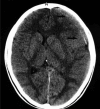
Case description: A 15-year-old healthy boy with a recent history of head trauma experienced headache that gradually worsened over the course of 10 days. He underwent CT and MRI brain scans which revealed the presence of subdural haematoma caused by the rupture of a middle cranial fossa arachnoid cyst. This was accompanied by intracystic haemorrhage. The subdural haematoma was removed, while communication of the cyst with the basal cisterns was also performed. The postoperative course of the patient was uneventful.
Conclusions: The annual haemorrhage risk for the patients with middle cranial fossa cysts remains very low. However, when haemorrhage occurs, in most occasions it can be effectively managed only with haematoma evacuation.
Keywords: intracranial arachnoid cyst; intracystic bleeding; subdural haematoma; treatment.
Figures


References
-
- Iaconetta G, Esposito M, Maiuri F, Cappabianca P. Arachnoid cyst with intracystic haemorrhage and subdural haematoma: case report and literature review. Neurol Sci. 2006;26:451–455. - PubMed
-
- Gosalakkal JA. Intracranial arachnoid cysts in children: a review of pathogenesis, clinical features and management. Pediatr Neurol. 2002;26:93–98. - PubMed
-
- Hopkin J, Mamourian A, Lollis S, Duhaime T. The next extreme sport? Subdural haematoma in a patient with arachnoid cyst after head shaking competition. Br J Neurosurg. 2006;20:111–113. - PubMed
-
- Parsch CS, Krauss J, Hofmann E, Meixensberger J, Roosen K. Arachnoid cysts associated with subdural hematomas and hygromas: analysis of 16 cases, long-term follow-up and review of the literature. Neurosurgery. 1997;40:483–490. - PubMed
-
- Kanev PM. Chapter208, Arachnoid cyst. In: Youmans JR, editor. Neurological Surgery. ed 3. vol 3. Philadelphia: Saunders; 2004. pp. 3289–3299.
Publication types
LinkOut - more resources
Full Text Sources
