. 2008 Dec;15(12):3538-49.
doi: 10.1245/s10434-008-0046-4. Epub 2008 Oct 16.

Evaluation of the sentinel immunized node for immune monitoring of cancer vaccines

Affiliations

Evaluation of the sentinel immunized node for immune monitoring of cancer vaccines

Craig L Slingluff Jr et al. Ann Surg Oncol. 2008 Dec.

Abstract

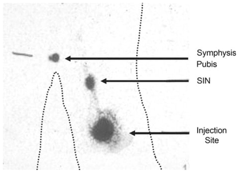
Background: We hypothesized that lymph nodes draining sites of cutaneous vaccination could be identified by sentinel node biopsy techniques, and that measuring T-cell response with lymphocytes obtained from these lymph nodes would provide a more sensitive measure of immunogenicity than would the same measurement made with peripheral blood lymphocytes (PBL).

Methods: ELISpot analysis was used to determine the magnitude of vaccine-specific T-cell response in the sentinel immunized nodes (SIN), random lymph nodes, and peripheral blood lymphocytes (PBL) obtained from patients enrolled in clinical trials of experimental melanoma vaccines.

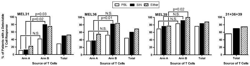
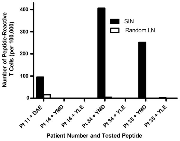
Results: The SIN biopsy was successful in 97% of cases and morbidity was very low. The T-cell response to vaccination was detected with greater sensitivity in the SIN (57%) than in PBL (39%), and evaluation of T-cell responses in the SIN and the PBL together yielded T-cell responses in 63% of patients. When the T-cell responses from a SIN and a random lymph node were compared in four patients, immune responses were detected to one of the vaccine peptides in three of these four patients. In all of those cases, responses were present in the SIN but absent from the random lymph node.

Conclusion: Measurements of T-cell responsiveness to cutaneous immunization are more frequently positive in the SIN than they are in the PBL, however evaluation of both the SIN and PBL permit a more sensitive measure of T-cell immunogenicity than use of either single source.


Figures

FIG. 1
FIG. 1
Identification of the SIN in the groin draining a cutaneous vaccine site in the thigh by Tc99-sulfur colloid and excision guided by a gamma probe. Technetium-labeled sulfur colloid was injected intradermally around the vaccine injection site reaction in the left thigh. The resulting gamma camera scan demonstrates the radio-activity at the injection site, the lymphatic channel draining toward the sentinel immunized node, and the hot spot representing the location of the SIN in the left groin. The location of the symphysis pubis was identified with a radioactive marker. The body contours are marked with dotted lines.
FIG. 2
FIG. 2
Immunohistochemical identification and quantification of S100+ dendritic cells in two different SINs. A central slice of each immunized lymph node was preserved in formalin and evaluated by immunohistochemical stains for S100+ dendritic cells. The proportion of the cross-sectional area occupied by DC ranged from about 2% to 20%. Examples are shown for patient VMM226 (A, B) with 6.6% of the lymph node containing S100+ dendritic cells and for VMM193 (C, D) with 2.7% of the lymph node containing S100+ dendritic cells. Histologically, the lymph node from patient VMM226 (A) had reactive features, whereas the lymph node from patient VMM193 (B) did not have reactive features. (A) and (C) were at an original magnification of 20× with the boxed areas approximately corresponding to (B) and (D); (B) and (D) were at an original magnification of 100×.
FIG. 3
FIG. 3
Patients on the three vaccine trials Mel31, Mel36, and Mel39 were evaluated for evidence of T-cell response in the PBL and in the SIN. The proportion of patients with T-cell responses to at least one peptide in the PBL, in the SIN, or in either the PBL or SIN are presented from the individual trials as well as for the three trials combined (N = 110). The Mel31, Mel36, and Mel37 trials each had two study arms (A and B) which are described in the “Patients and Methods” section. Results are reported for the individual arms of each study, for each study as a whole (“Total”), and the combined results of all three studies (“31 + 36 + 39”). Statistical significance was determined with Fisher’s exact test. N.S., not significant.
FIG. 4
FIG. 4
T-cell response in SIN is specific, and is not evident in a random node from the same basin (Mel36). Lymphocytes from a SIN and from a random, nonradioactive node were sensitized in vitro once with the four melanoma peptide mixture, then assayed by ELIspot assay for reactivity to each of the peptides relevant for the patient’s HLA type. Patients 11, 14, 34, and 35 were studied. Patient 11 was HLA-A1+ and reactivity was tested to the tyrosinase peptide DAEKSDICTDEY. The other three patients were HLA-A2+ and reactivity was tested to the gp100 and tyrosinase peptides YLEPGPVTA and YMDGTMSQV. The reactivity from the SIN is shown in the solid black bars. Reactivity from the random nodes is shown in the white empty bars. Reactivity was negative for patient 14, and reactivity to YLEPGPVTA was negative in all three cases evaluated.
FIG. 5
FIG. 5
SIN size and cell yield by clinical trial study arm. Mel31-A and Mel31-B are two arms of the same study, while Mel36-A and Mel36-B are two arms of a second study and Mel39-A and Mel39-B are two of a third study. The Mel37 trial did not have separate arms. (A) The mean calculated volume of the SIN±1 standard deviation (SD). (B) The mean number of cells obtained from each SIN±1 SD for patients on each arm of the trials are plotted as open bars. The Mann–Whitney test was used to determine statistical significance. N.S., not significant.

References

    1. Rosenberg SA, Yang JC, Restifo NP. Cancer immunotherapy: moving beyond current vaccines. Nat Med. 2004;10:909–15. - PMC - PubMed
    1. Drake CG, Jaffee E, Pardoll DM. Mechanisms of immune evasion by tumors. Adv Immunol. 2006;90:51–81. - PubMed
    1. Slingluff CL, Chianese-Bullock KA, Bullock TNJ, et al. Immunity to melanoma antigens: from self-tolerance to immunotherapy. Adv Immunol. 2006;90:243–95. - PubMed
    1. Marchand M, van Baren N, Weynants P, et al. Tumor regressions observed in patients with metastatic melanoma treated with an antigenic peptide encoded by gene MAGE-3 and presented by HLA-A1. Int J Cancer. 1999;80:219–30. - PubMed
    1. Coulie PG, Karanikas V, Colau D, et al. A monoclonal cytolytic T-lymphocyte response observed in a melanoma patient vaccinated with a tumor-specific antigenic peptide encoded by gene MAGE-3. Proc Natl Acad Sci USA. 2001;98:10290–5. - PMC - PubMed

Publication types