. 2008 Nov;237(11):3270-9.
doi: 10.1002/dvdy.21740.

Biphasic Ngn3 expression in the developing pancreas

Affiliations

Biphasic Ngn3 expression in the developing pancreas

Alethia Villasenor et al. Dev Dyn. 2008 Nov.

Abstract

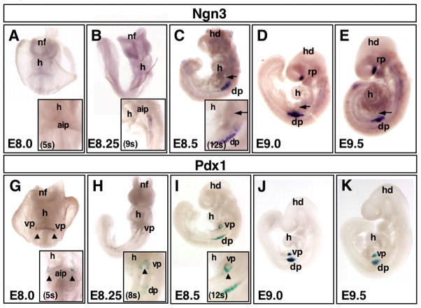
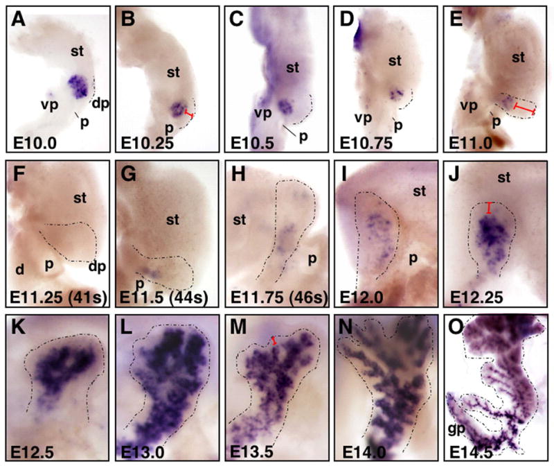
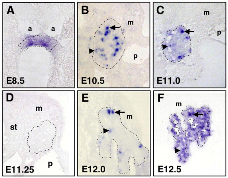
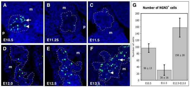
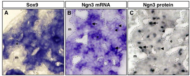
Ngn3 is a bHLH transcription factor critical for the specification of endocrine cells in the pancreatic Islets of Langerhans. Previous studies in mouse embryos have reported transient expression of Ngn3 in scattered cells within the developing pancreatic epithelium during midgestation (Schwitzgebel et al. [2000] Development 127:3533-3542). Specifically, these Ngn3-expressing cells have been shown to be progenitor cells fated to give rise to islet endocrine cells (Gradwohl et al. [2000] Proc Natl Acad Sci USA 97:1607-1611). Here, we characterize the expression of Ngn3 transcripts and protein throughout pancreatic development. Interestingly, we identify and define a dramatic and previously unnoticed gap in developmental Ngn3 expression. We show that both Ngn3 transcript and protein expression occur in two distinct temporal waves, the first occurring early from approximately E8.5 to E11.0, and the second initiating at approximately E12.0. Strikingly, this observed biphasic expression correlates with the "first" and "second" transitions, which encompass two distinct waves of embryonic endocrine differentiation. In addition, our studies demonstrate that Ngn3 transcripts are markedly more widespread in the pancreatic epithelium than NGN3 protein, indicating that post-transcriptional regulation is likely to play a critical role during endocrine differentiation.


Figures

Figure 1
Figure 1. Initiation of Ngn3 and Pdx1 expression during embryogenesis
Developmental profile comparison of early expression of Ngn3 and Pdx1, using whole mount in situ hybridization of Ngn3 (A–E) and whole mount β-galactosidase staining of Pdx1-lacZ embryos (G–K) from E8.0 to E9.5. Embryos in A,B,G,H face forward. Embryos in C–E and I–K face left. Ngn3 and Pdx1 initiate in the dorsal pancreas at relatively similar timepoints shortly after turning, between E8.25 and E8.5 (A–C Ngn3, G–I Pdx1). Ngn3 initiates in the dorsal pancreas at 9s (inset in B). Pdx1 is expressed in two ventral pancreas domains, as early as E8.0 (5s) (arrowheads in G). These ventral domains fuse as the embryo turns (arrowheads in H,I). While Pdx1 marks the ventral pancreas from early stages (G–K), Ngn3 is not expressed in the ventral pancreas (arrows in C–E) at any time during early development (prior to E10.0). See insets in (A–C) for close-ups on early dorsal and ventral domains for onset of Ngn3 expression, and insets in (G–I) for close-ups of Pdx1 initiation. Note also that Ngn3 marks anterior neurectoderm in the forebrain (head staining in D,E), ventral to Rathke’s pouch. dp, dorsal pancreatic bud; h, heart; hd, head; nf, neural folds; rp, Rathke’s pouch; vp, ventral pancreatic bud.
Figure 2
Figure 2. Biphasic transcription of Ngn3 in the developing pancreas
Whole mount in situ hybridization of Ngn3 in wildtype embryos from E10.0 to E14.5. A–G) Dissected gut tubes, anterior is up, dorsal is to the right; H) gut tube in process of turning, anterior is up, but pancreas is shifting relative to stomach, and focus is on pancreas near portal vein; I–O) Dorsal pancreatic bud, with stomach seen in background, except in O where pancreas is isolated. Dorsal pancreas in all panels is outlined with a black dotted line. A–C) Note robust expression of Ngn3 in dorsal pancreatic bud at E10.0 to E10.5. D,E) Decline of Ngn3 expression begins at E10.75, and F) is undetectable at E11.25. G–K) Expression then begins to increase from E11.5–12.5, after which expression is strong in cells of the branching epithelium (L–O). A–E) Note that Ngn3 expression in ventral pancreas is low at all stages shown. Ventral pancreas is not shown in (F–O). After E11.0, the ventral pancreas grows posteriorly, along the mesentery of the duodenum, and is not visible in images as presented. Note also that distal pancreatic mesenchyme (or ‘cap’) is thickest around E11.0, but becomes progressively thinner as development proceeds (compare red brackets in B,E,J,M). Precise staging of embryos was accomplished by counting somites (see Table 1). dp, dorsal pancreatic bud; d, duodenum; gp, gastric pancreas; p, portal vein; st, stomach; vp, ventral pancreatic bud.
Figure 3
Figure 3. Ngn3 expression in pancreatic epithelium
Sections showing in situ hybridization of Ngn3 in wildtype embryos from E8.5 to E12.5. A) Note more homogeneous expression in early endoderm (E8.5). At this stage, the endoderm has not yet thickened or stratified and is still a cuboidal epithelium. B) At E10.5, expression becomes heterogeneous with ‘high’ (arrow) and ‘low’ (arrowhead) expressing cells. C) By E11.0, fewer scattered cells still express higher levels of Ngn3 (arrow), while expression levels decline in the rest of the epithelium (arrowhead). D) Ngn3 expression is undetectable at E11.25. E) Cells expressing Ngn3 can be easily detected at E12.0. F) Expression then increases, both at higher levels in scattered cells and at lower levels throughout the epithelium (E12.5 shown). Pancreatic epithelium is outlined with a dotted line. a, aortae; m, mesenchyme; p, portal vein; st, stomach.
Figure 4
Figure 4. Biphasic NGN3 expression in pancreatic epithelium
NGN3 protein detected by immunofluorescence on sectioned pancreas, from E10.5 to E13.5. A) NGN3 protein is readily detected in the stratified epithelium of the E10.5 bud. B,C) From E11.25 to E11.5, few if any cells show NGN3 staining in single sections. D–F) At E12.0, we start to detect a steady increase in the number of cells expressing NGN3 that continues to E13.5. Pancreatic epithelium is outlined with a white dotted line. High (arrows) and low (arrowheads) levels of NGN3 protein can be observed in individual cells. m, mesenchyme; p, portal vein. G) Quantification of the number of cells showing NGN3 protein expression at E10.5, E11.5 and E12.5–E13.0 (n=3 for each stage). The average number of cells showing protein expression at E11.5 is approximately 30, while at E10.5 it is 96 and between E12.5–E13.5 it is 158. Note the statistiscally significant reduction of NGN3+ cells at E11.5 (p≤0.02).
Figure 5
Figure 5. Ngn3 transcripts are more widespread in pancreatic epithelium than NGN3 protein
Sections through E13.0 dorsal pancreas. A,B) In situ hybridzation. C) Immunohistochemistry for NGN3. A–C) High magnification of the pancreatic epithelium (20X). A) Sox9 is expressed throughout the pancreatic epithelium. B) Ngn3 is expressed in many cells within the pancreatic epithelium, displaying both high (arrows) and low (arrowheads) expressing cells. C) NGN3 protein is concentrated in scattered cells of the pancreatic epithelium. In addition, cells are observed that express either high (arrows) or low (arrowheads) levels of NGN3 protein. Note that there are significantly more cells that express the Ngn3 transcript (B), than cells that express NGN3 protein (C). Pancreatic epithelium is outlined with a dotted line. e, epithelium; m, mesenchyme.

References

    1. Ahlgren U, Jonsson J, Edlund H. The morphogenesis of the pancreatic mesenchyme is uncoupled from that of the pancreatic epithelium in IPF1/PDX1-deficient mice. Development. 1996;122:1409–1416. - PubMed
    1. Apelqvist A, Li H, Sommer L, Beatus P, Anderson DJ, Honjo T, Hrabe de Angelis M, Lendahl U, Edlund H. Notch signalling controls pancreatic cell differentiation. Nature. 1999;400:877–881. - PubMed
    1. Artner I, Blanchi B, Raum JC, Guo M, Kaneko T, Cordes S, Sieweke M, Stein R. MafB is required for islet beta cell maturation. Proc Natl Acad Sci U S A. 2007;104:3853–3858. - PMC - PubMed
    1. Beaupain D, Dieterlen-Lievre F. An immunocytological study of differentiation of the endocrine pancreas of the chick embryo. II. Glucagon. Gen Comp Endocrinol. 1974;23:421–431. - PubMed
    1. Burlison JS, Long Q, Fujitani Y, Wright CV, Magnuson MA. Pdx-1 and Ptf1a concurrently determine fate specification of pancreatic multipotent progenitor cells. Dev Biol. 2008;316:74–86. - PMC - PubMed

Publication types

MeSH terms

Substances

LinkOut - more resources