. 2008 Nov;118(11):3671-81.
doi: 10.1172/JCI36942. Epub 2008 Oct 16.

Human sperm devoid of PLC, zeta 1 fail to induce Ca(2+) release and are unable to initiate the first step of embryo development

Affiliations

Human sperm devoid of PLC, zeta 1 fail to induce Ca(2+) release and are unable to initiate the first step of embryo development

Sook-Young Yoon et al. J Clin Invest. 2008 Nov.

Abstract

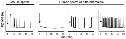
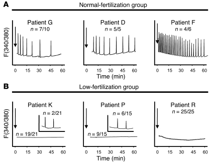
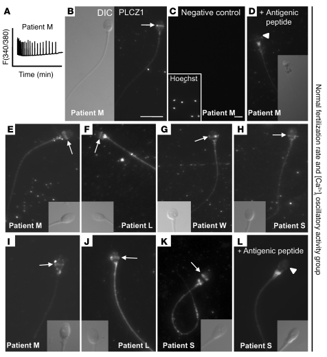
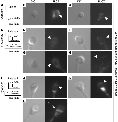
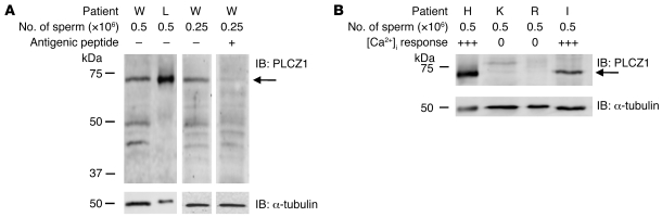
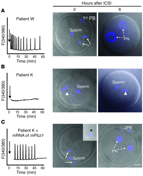
Egg activation, which is the first step in the initiation of embryo development, involves both completion of meiosis and progression into mitotic cycles. In mammals, the fertilizing sperm delivers the activating signal, which consists of oscillations in free cytosolic Ca(2+) concentration ([Ca(2+)](i)). Intracytoplasmic sperm injection (ICSI) is a technique that in vitro fertilization clinics use to treat a myriad of male factor infertility cases. Importantly, some patients who repeatedly fail ICSI also fail to induce egg activation and are, therefore, sterile. Here, we have found that sperm from patients who repeatedly failed ICSI were unable to induce [Ca(2+)](i) oscillations in mouse eggs. We have also shown that PLC, zeta 1 (PLCZ1), the sperm protein thought to induce [Ca(2+)](i) oscillations, was localized to the equatorial region of wild-type sperm heads but was undetectable in sperm from patients who had failed ICSI. The absence of PLCZ1 in these patients was further confirmed by Western blot, although genomic sequencing failed to reveal conclusive PLCZ1 mutations. Using mouse eggs, we reproduced the failure of sperm from these patients to induce egg activation and rescued it by injection of mouse Plcz1 mRNA. Together, our results indicate that the inability of human sperm to initiate [Ca(2+)](i) oscillations leads to failure of egg activation and sterility and that abnormal PLCZ1 expression underlies this functional defect.


Figures

Figure 1
Figure 1. Injection of mouse and human sperm into mouse eggs induces [Ca2+]i oscillations.
Mouse sperm (left panel) induce consistent oscillations (representative of at least 10 sperm per male and of at least 10 different males), whereas human sperm induce highly variable responses according to male of origin (3 right panels). Each panel corresponds to a different patient and is representative of the activity of the sperm for the particular male. Arrows denote the time of sperm injection. [Ca2+]i monitoring commenced within 10 minutes of injection. These experiments were repeated at least 5 times. F, fluorescence ratio.
Figure 2
Figure 2. Sperm from patients with different ICSI fertilization rates initiate widely dissimilar [Ca2+]i oscillations in mouse eggs.
(A) [Ca2+]i profiles of 3 patients with high fertilization rates are shown. (B) Profiles belong to 3 patients who failed or showed low fertilization rates following ICSI. Insets in panels for patients K and P depict representative patterns of [Ca2+]i responses observed in the few eggs that initiated oscillations following sperm injection. Arrows denote time of sperm injection. n, numerator indicates the number of eggs that displayed the [Ca2+]i profile shown, whereas denominator denotes the total number of eggs tested.
Figure 3
Figure 3. PLCZ1 localizes to the equatorial/postacrosomal region of human sperm.
Two different antibodies, MI-305 (BH) and PF-354 (IL), and sperm from different patients capable of inducing [Ca2+]i oscillations were used to characterize PLCZ1 localization. In BD, localization of PLCZ1 and specificity of the MI-305 antibody were characterized using patient M’s sperm, which induced high-frequency oscillations (A). (B) Left panel shows bright field image, whereas right panel shows the corresponding immunofluorescent image. Arrows denote PLCZ1 localization. Negative controls were incubated in the absence of primary antibody (C) or after incubation with antigenic peptide (D). (C) Inset shows Hoechst 33342–stained sperm nuclei. Original magnification, ×630. (D) Inset shows reduced (50%) bright field image of fluorescent image in panel. (D) White arrowheads denote the loss of the PLCZ1 band in the presence of antigenic peptide. (EH) Staining of different patients’ sperm (insets show corresponding bright field images). (IL) Staining of same patients’ sperm but using the PF-354 antibody. Insets show corresponding bright field images. Scale bars: 10 μm. Scale bar for B also applies to parts DL.
Figure 4
Figure 4. Reduced expression of PLCΖ1 in the sperm of patients who fail ICSI and lack [Ca2+]i oscillatory ability.
MI-305 antibody and sperm from 3 patients who failed ICSI and lacked [Ca2+]i oscillatory activity (A, D, and I) were used to examine PLCZ1 localization. (B and C) Left panels show bright field images of sperm from patient R; right panels show the corresponding immunofluorescent images. White arrowheads point to the expected location of PLCZ1. (EH and JL) Sperm from patients K and P were examined, respectively, using the MI-305 antibody. (L) Arrow denotes PLCZ1 localization. Scale bar: 10 μm.
Figure 5
Figure 5. Expression of PLCZ1 by IB in sperm of patients capable of inducing [Ca2+]i oscillations and/or fertilization and in sperm of patients who failed ICSI.
(A) Band of approximately 70 kDa was detected in sperm extracts of patients whose sperm showed [Ca2+]i oscillatory activity; arrow on the right side of the panel denotes the expected MW of PLCZ1. The intensity of the 70-kDa band was reduced by preincubation of the MI-305 antibody with the appropriate antigenic peptide. (B) Expression of PLCΖ1 was detected in the extracts of 2 other patients with successful ICSI outcomes, but it was absent in patients K and R, who failed ICSI. IB of α-tubulin was used as a loading control and shows equal loading per lane.
Figure 6
Figure 6. Injection of mouse Plcz1 mRNA rescues egg activation failure after ICSI in mouse eggs.
(A) Injection of a sperm from patient W, with normal [Ca2+]i oscillatory activity (left panel), induces resumption of meiosis after 2 hours (arrows denote anaphase II) and PN formation (broken arrows) by 6 hours after injection. (B) Injection of sperm from patient K, which is unable to initiate [Ca2+]i oscillations (left panel), fails to induce egg activation by either 2 hours or 6 hours (arrowhead denotes MII chromatin). (C) Injection of mouse Plcz1 mRNA (0.1 μg/μl) 2 hours after injection of sperm initiates fertilization-like oscillations and rescues egg activation, as shown by the resumption of meiosis (2 hours) and PN formation (6 hours; 8 hours after injection of patient K’s sperm). 1st PB, first polar body; 2PB, second polar body. TO-PRO-3 staining (blue) was used to stain the chromatin. Asterisk in inset points to the persistence of the human sperm tail in mouse eggs. Scale bar: 10 μm.

References

    1. Schultz R.M., Kopf G.S. Molecular basis of mammalian egg activation. Curr. Top. Dev. Biol. 1995;30:21–62. doi: 10.1016/S0070-2153(08)60563-3. - DOI - PubMed
    1. Kline D., Kline J.T. Repetitive calcium transients and the role of calcium in exocytosis and cell cycle activation in the mouse egg. Dev. Biol. 1992;149:80–89. doi: 10.1016/0012-1606(92)90265-I. - DOI - PubMed
    1. Miyazaki S., et al. Temporal and spatial dynamics of the periodic increase in intracellular free calcium at fertilization of golden hamster eggs. Dev. Biol. 1986;118:259–267. doi: 10.1016/0012-1606(86)90093-X. - DOI - PubMed
    1. Stricker S.A. Comparative biology of calcium signaling during fertilization and egg activation in animals. Dev. Biol. 1999;211:157–176. doi: 10.1006/dbio.1999.9340. - DOI - PubMed
    1. Miyazaki S., Ito M. Calcium signals for egg activation in mammals. J. Pharmacol. Sci. 2006;100:545–552. doi: 10.1254/jphs.CPJ06003X. - DOI - PubMed

Publication types

MeSH terms