. 2008 Nov;38(11):3167-77.
doi: 10.1002/eji.200838456.

Migration of antibody secreting cells towards CXCL12 depends on the isotype that forms the BCR

Affiliations

Migration of antibody secreting cells towards CXCL12 depends on the isotype that forms the BCR

Gertrude Achatz-Straussberger et al. Eur J Immunol. 2008 Nov.

Abstract

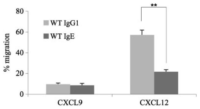
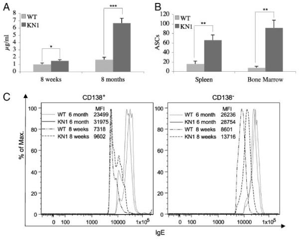
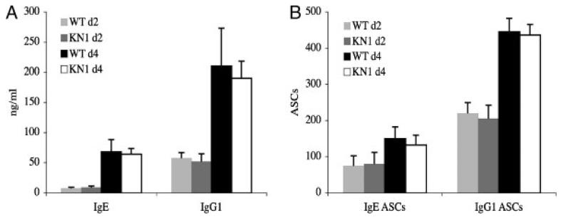
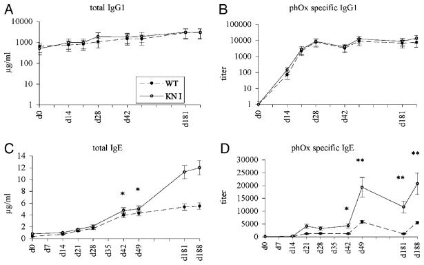
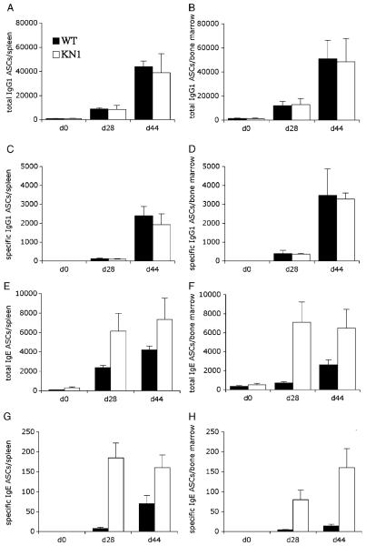
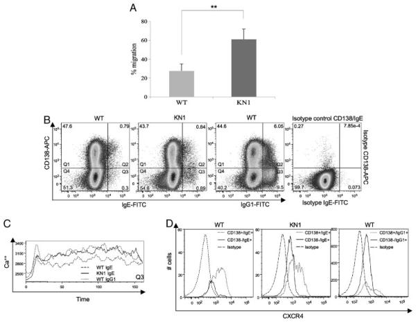
Truncation of the cytoplasmic tail of membrane-bound IgE in vivo results in lower serum IgE levels, decreased numbers of IgE-secreting plasma cells and the abrogation of specific secondary immune responses. Here we present mouse strain KN1 that expresses a chimeric epsilon-gamma1 BCR, consisting of the extracellular domains of the epsilon gene and the transmembrane and cytoplasmic domains of the gamma1 gene. Thus, differences in the IgE immune response of KN1 mice reflect the influence of the "gamma1-mediated signalling" of mIgE bearing B cells. KN1 mice show an increased serum IgE level, resulting from an elevated number of IgE-secreting cells. Although the primary IgE immune response in KN1 mice is inconspicuous, the secondary response is far more robust. Most strikingly, IgE-antibody secreting cells with "gamma1-signalling history" migrate more efficiently towards the chemokine CXCL12, which guides plasmablasts to plasma cell niches, than IgE-antibody secreting cells with WT "epsilon-signalling history". We conclude that IgE plasmablasts have an intrinsic, lower chance to contribute to the long-lived plasma cell pool than IgG1 plasmablasts.


Figures

Figure 1
Figure 1
Migration assay of IgE- and IgG1-ASC. IgG1-ASC and IgE-ASC were exposed to the chemokine CXCL9 and CXCL12 in a Transwell culture plate and their migratory behaviour was tested (see also Supporting Information Fig. 1). This figure summarizes data from an experiment repeated three times using three mice per group. Potentially migrating cells of each subclass were counted by ELISPOT and set as 100%. Using the paired t-test, statistical significance at the level of **p<0.001 was calculated.
Figure 2
Figure 2
IgE serum levels, ELISPOT and flow cytometric analysis of bone marrow and spleen of WT and KN1 mice. (A) Total serum IgE levels of five young (8 wk) and five old (8-months) WT and KN1 mice. (B) ELISPOT analysis of IgE-ASC originating from pooled spleen and bone marrow cells from five WT and five KN1 mice. For statistical analysis, IgE-ASC were determined individually and extrapolated for 106 spleen or bone marrow cells. Values are means±SD of five mice per time point. (A,B) Data from the described experiments repeated three times, five mice per group. (*p<0.05; **p<0.001; ***p<0.0001). (C) Flow cytometric analysis of bone marrow cells of 8 wk and 6-month-old WT and KN1 mice. Bone marrow cells were incubated with ice-cold stripping buffer followed by incubation with rat anti-CD138 and rat-anti IgE. Cells were pre-gated for living lymphocytes. The lower cutoff value for mIgE+ cells was determined with corresponding isotype controls (see Fig. 6B).
Figure 3
Figure 3
In vitro differentiation potential of activated IgM+ cells of WT and KN1 mice. In vitro activated IgM+ cells of WT and KN1 mice were cultured for 2 and 4 days with LPS and IL-4. (A) Supernatants were analysed for IgE and IgG1 content by ELISA. (B) Cells were analysed for IgE and IgG1 secretion by ELISPOT. Values for A and B are means±SD of three independent experiments with three mice each.
Figure 4
Figure 4
Time course of immunization I. Five WT and five KN1 mice were immunized with the T-cell dependent antigen phOx-OVA at days 0, 14, 21, 42 and 181. Immunoglobulin levels were measured by ELISA. (A) Total serum IgG1; (B) phOx-specific IgG1; (C) total IgE; (D) phOx-specific IgE. Values are means±SD of three immunizations with five mice per time point. p-values for time points d42, d49, d181 and d188 for total and specific IgE were significant (*p<0.05 and **p<0.001).
Figure 5
Figure 5
Time course of immunization II. Five WT and five KN1 mice were immunized with OVA. IgE- and IgG1-ASC of spleen and bone marrow were detected with ELISPOT assays. The total and specific humoral IgE response at day 28 of KN1 mice was significantly up-regulated in the spleen (E and G) and even more in the bone marrow (F and H). Corresponding measurement of IgG1-ASC (A–D) served as control. Values are means±SD of three immunization experiments with five mice per time point.
Figure 6
Figure 6
Transwell migration assay of WT- and KN1-derived IgE-ASC/evaluation of the surface co-expression of mIgE, CD138 and CXCR4 and functional activity of CD138low, mIgE(mIgG1)+ cells. (A) Summary of an experiment repeated three times, with three mice per group, investigating the migration frequency of IgE-ASC towards CXCL12. Values are means±SD (**p<0.001). (B) To visualize mIgE+ populations co-expressing CD138, 2 × 10⁶ activated lymphocytes were acquired. The lower cutoff value for mIgE+ cells was determined with corresponding isotype controls and competition stainings. Numbers in the quadrants represent % of the cell populations. (C) Ca2+ fluxes of CD138low, mIg+ B cells (see Quadrant Q3 of Fig. 6B). (D) CD138low, mIgE+ and CD138high, mIgE+ populations were gated and further analysed for CXCR4 expression.

References

    1. Manz RA, Hauser AE, Hiepe F, Radbruch A. Maintenance of serum antibody levels. Annu. Rev. Immunol. 2005;23:367–386. - PubMed
    1. McHeyzer-Williams MG. B cells as effectors. Curr. Opin. Immunol. 2003;15:354–361. - PubMed
    1. Radbruch A, Muehlinghaus G, Luger EO, Inamine A, Smith KG, Dorner T, Hiepe F. Competence and competition: the challenge of becoming a long-lived plasma cell. Nat. Rev. Immunol. 2006;6:741–750. - PubMed
    1. Calame KL. Plasma cells: finding new light at the end of B cell development. Nat. Immunol. 2001;2:1103–1108. - PubMed
    1. Manz RA, Lohning M, Cassese G, Thiel A, Radbruch A. Survival of long-lived plasma cells is independent of antigen. Int. Immunol. 1998;10:1703–1711. - PubMed

Publication types

LinkOut - more resources