Comparative Study
. 2008 Dec 20;511(6):788-803.
doi: 10.1002/cne.21867.

Tau isoform regulation is region- and cell-specific in mouse brain

Affiliations
Comparative Study

Tau isoform regulation is region- and cell-specific in mouse brain

Pamela McMillan et al. J Comp Neurol. .

Abstract

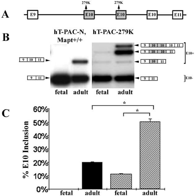
Tau is a microtubule-associated protein implicated in neurodegenerative tauopathies. Alternative splicing of the tau gene (MAPT) generates six tau isoforms, distinguishable by the exclusion or inclusion of a repeat region of exon 10, which are referred to as 3-repeat (3R) and 4-repeat (4R) tau, respectively. We developed transgenic mouse models that express the entire human MAPT gene in the presence and absence of the mouse Mapt gene and compared the expression and regulation of mouse and human tau isoforms during development and in the young adult. We found differences between mouse and human tau in the regulation of exon 10 inclusion. Despite these differences, the isoform splicing pattern seen in normal human brain is replicated in our mouse models. In addition, we found that all tau, both in the neonate and young adult, is phosphorylated. We also examined the normal anatomic distribution of mouse and human tau isoforms in mouse brain. We observed developmental and species-specific variations in the expression of 3R- and 4R-tau within the frontal cortex and hippocampus. In addition, there were differences in the cellular distribution of the isoforms. Mice transgenic for the human MAPT gene exhibited higher levels of neuronal cell body expression of tau compared to wildtype mice. This neuronal cell body expression of tau was limited to the 3R isoform, whereas expression of 4R-tau was more "synaptic like," with granular staining of neuropil rather than in neuronal cell bodies. These developmental and species-specific differences in the regulation and distribution of tau isoforms may be important to the understanding of normal and pathologic tau isoform expression.


Figures

Figure 1
Figure 1
Human MAPT and mouse Mapt RNA levels in wildtype mice and in mice transgenic for human MAPT (hT-PAC-N Mapt+/+). RNA from either mouse or human brain was isolated as described in the Materials and Methods section and RT-PCR was performed using either mouse or human-specific primers that amplified transcripts from mid-E9 to mid-E11. The short fragment (165 bp) is E10- and the long fragment (265 bp) is E10+. A. Mouse-specific RT-PCR primers amplify mouse Mapt RNA but not human MAPT RNA. Using mouse-specific primers (ME9F2 and ME11R), no transcript was detected in human embryonic (E14) or adult brain RNA. In fetal mice (E14) the predominant product is E10- although some E10+ RNA is detected. In RNA from adult (90 days) hT-PAC-N, Mapt+/+ lines that have an intact mouse Mapt gene, the predominant transcript from the mouse gene is E10+. B. Human-specific RT-PCR primers amplify only transcripts from the human gene but not the mouse gene. Samples are the same as in A and the primers used were HE9F2 and HE11R. C. Quantitation of E10+ transcripts using mouse-specific and human-specific primers to compare the appearance of E10+ mouse Mapt and human MAPT transcripts in developing transgenic mouse brains. For WT mice (black bars), each bar represents values from 2 to 5 mice. For hT-PAC-N, Mapt+/+, each bar for mouse (gray) and human (open) is the average combined E10+ value from mouse lines 9, 12 and 13 and represents a minimum of 6 to a maximum of 14 mice. Mouse Mapt transcripts are predominantly E10- until P6 when the amount of E10+ begins to increase (WT 79.7% vs. transgenic 75.8%) until by day P24, all Mapt transcripts are E10+. The presence of the human MAPT gene in hT-PAC-N, Mapt+/+ does not influence the endogenous pattern of E10 splicing of transcripts from the mouse Mapt gene compared with non-transgenic mice. The human gene in the mouse produces no detectable E10+ transcripts until P12 (14.01%), at which stage endogenous mouse E10+ levels are significantly higher at 76% than human E10+ (p < 10-16). D. In adult human brain, transcripts from MAPT are 49 % E10+ (SD 0.03). For the human gene in mice (hT-PAC-N, mapt +/+), 21 % of transcripts were E10+ (SD 1.2, n = 13). This is the average from 4 different hT-PAC-N Mapt+/+ lines in adult (P90) mice with an intact Mapt gene. Error bars for individual lines represent values from at least 3 different brain samples with standard deviations as follows: adult human brain (SD = 3.0, n = 3), transgenic mice Line 12 (SD = 1.0, n = 3), Line 9 (SD = 1.0, n = 3), Line 4 (SD = 0.6, n = 4) and Line 13 (SD = 0.1, n = 13).
Figure 2
Figure 2
Immunoblot analysis of RAB soluble human MAPT and mouse Mapt isoforms in brains from wildtype and transgenic mice. Whole brain homogenates were prepared as described in the Materials and Methods and subjected to electrophoresis and immunoblot analysis using the following antibodies: RD4 (1-1000 dilution, top panels, A and B); RD3 (1-3000 dilution, middle panels, A and B); Rb17025 (diluted 1-6000, bottom panel, A and B) which recognizes both mouse and human tau (all six isoforms); T14 (1-1000) (C, all panels), an antibody specific for human tau (all six isoforms) that does not recognize mouse tau. Protein loads/lane for A and B are: 1.2 ug, top panel; 0.1 ug, middle panel; 0.2 ug, bottom panel. Lanes labeled with “-” are untreated soluble tau and lanes labeled “+” were treated with λ-phosphatase. A. Wildtype mice produce predominantly 3R tau at P6, both 3R and 4R tau at P24 and predominantly 4R tau at P90. Note that for all isoforms, the mouse protein migrates faster than the human equivalent protein. However, spacing is not altered. B. The human gene in a Mapt-/- mouse produces predominantly 3R tau and no 4R tau at P6. By P24, other tau isoforms were observed including some that are 4R, although the predominant isoform is still 3R even in the adult mice (P90). C. The human gene in the adult mouse brain produces all 6 tau isoforms and FTDP-17 mutations that alter isoform ratios in man also affect splicing of E10 in mouse. RAB soluble whole brain homogenates were prepared from mice transgenic for either the normal MAPT gene (hT-PAC-N) or MAPT with the FTDP-17 mutations V337M, R5L, S305S, E10+14, P301L, or N279K on a mouse Mapt+ background. Samples in the top panel were untreated and samples in the bottom panel were treated with λ-phosphatase. Since different transgenic lines produce different amounts of human tau, loading was adjusted so that approximately the same amount of human tau was present in each lane. The protein loaded for phosphatase treated samples was as follows: 0.25 ug, hT-PAC-N; 0.17 ug, hT-PAC-337M; 0.43 ug, hT-PAC-5L; 0.9 ug, hT-PAC-305S; 0.18 ug, hT-PAC-E10+14; 0.2 ug, hT-PAC-301L; and 0.2 ug, hT-PAC-279K. The protein loaded for untreated samples was 0.5 ug, except for hT-PAC-337M, which was 0.1 ug. The standards are recombinant human tau.
Figure 3
Figure 3
The hT-PAC-279K transgene has 3 copies of E10 and the 279K mutation increases inclusion of E10. A. The structure of the E10 region of MAPT in mouse line hT-PAC-279K retaining 2 mutant E10 copies (shaded box) upstream of a normal E10 copy. B. RT-PCR products from fetal (E14) and adult (P90) hT-PAC-N Mapt+/+ and hT-PAC-279K mice. The structure for the RT-PCR products with multiple E10 copies was determined by DNA sequencing. Note that the normal E10 copy is spliced only in adult mouse brain and, in hT-PAC-279K, is detected as a minor species in the presence of both mutant E10 copies. C. Quantitation of E10 inclusion. Each bar represents values from 3 different brain samples. The 279K mutation in mice having multiple copies of E10, stimulates inclusion of one mutant E10 copy in fetal brain at 11.2 % (SD 4.8, n = 3) and predominantly enhances mutant E10+ transcripts in adult brain to 50.4 % (SD = 1.8, n = 2). The E10+ bar for hT-PAC-279K mice is the sum of all E10+ species as observed in B. A corrected significance criteria of p < 0.025 was used in E10 splicing comparisons between adult WT and mutant 279K mice as well as between fetal and adult 279K mice, p < 1 × 10⁻⁴. Solid bars, hT-PAC-N mice; hatched bars, hT-PAC-279K mice.
Figure 4
Figure 4
Expression of mouse 3R and 4R tau in the frontal cortex and hippocampus of wildtype mice at P3 (A, D, G, J), P6 (B, E, H, K), and P24 (C, F, I, L). In the cortex, 3R tau is expressed in all layers at all ages examined (A-C) while 4R tau is undetectable in P3 mice (D) but is observed beginning at P6 (E). Higher magnification (A’) of boxed region in A showing neuronal cell body staining of 3R tau at P3. In the hippocampus, little specific staining is seen at P3 for either 3R or 4R tau (G and J, respectively). At P6, 4R staining is seen in the hilus, stratum oriens and stratum radiatum (K), while 3R staining is still faint (H). By P24, both 3R and 4R are abundant throughout the hippocampus (I and L). Panels A-C and G-I were stained with the RD3 antibody and panels D-F and J-L were stained with the RD4 antibody. Scale bars A-L, 100 μm; A’, 25 μm. Abbreviations: Or (stratum oriens layer); Rad (stratum radiatum layer); MF (mossy fibers).
Figure 5
Figure 5
Expression of mouse tau isoforms and doublecortin (DCX) in the hippocampus of P24 wildtype mice. In the hippocampus of P24 wildtype mice, 3R tau (A, RD3 antibody) is expressed in the subgranular zone (SGZ) and 4R tau (B, RD4 antibody) is extensively expressed in the hilus and the inner dentate molecular layer (iDML). Mouse 3R tau and DCX are co-expressed in the same population of SGZ cells in the dentate gyrus of P24 wildtype mice (C, D). DCX was detected using an avidin-biotin complex and visualized with DAB under light microscopy (C). In the same tissue section, 3R tau was detected with an alkaline phosphatase avidin-biotin complex and visualized with Vector Red under immunofluoresence microscopy (D). Dcx and 3R tau are co-expressed in the same cells (E-G). DCX was detected using a Cy2-conjugated donkey anti-goat secondary antibody (E). 3R-tau was detected using a biotinylated M.O.M. mouse secondary antibody and the fluorescently tagged avidin system, Texas Red avidin D (F). Merge of the Cy-2 and Texas Red avidin D fluorescent signals (G). Arrowheads show individual cells within the SGZ that are co-labeled with both DCX and 3R-tau. Scale bars A-D, 100 μm; E-G, 25 μm. A magenta-green copy is available online (supplemental figure 2). Abbreviations: granular cell layer (GCL), dentate molecular layer (DML).
Figure 6
Figure 6
3R and 4R tau are undetectable in brain tissue of tau knockout mice. No immunostaining is observed in the hippocampus of P24 Mapt-/- mice using antibodies specific for 3R tau (RD3, A) and 4R tau (RD4, B). Scale bars A and B, 100 μm.
Figure 7
Figure 7
Expression of human 3R and 4R tau in the frontal cortex and hippocampus of hT-PAC-N, Mapt-/- mice at P3 (A, D, G, J), P6 (B, E, H, K), and P24 (C, F, I, L). In the cortex, 3R tau is observed in all cortical layers at all ages (A-C), while 4R tau is not observed at any age (D-F). Higher magnification (A’) of boxed region in A showing neuronal cell body staining of 3R tau at P3. In the hippocampus, 3R tau was expressed at all ages (G-I) while 4R tau was not detected until P24 (L). The transgenic animals expressed 3R tau in CA3 pyramidal neurons of the hippocampus (N). This neuronal cell body staining is also evident in human CA3 neurons (O) but is undetectable in the CA3 neurons of wildtype mice (M). Panels A-C, G-I and M-O were stained with the RD3 antibody and panels D-F and J-L were stained with the RD4 antibody. Scale bars A-O, 100 μm; A’, 25 μm. Abbreviations: MF (mossy fibers), CA3 (pyramidal neurons of the CA3).

References

    1. Andorfer C, Kress Y, Espinoza M, de Silva R, Tucker KL, Barde YA, Duff K, Davies P. Hyperphosphorylation and aggregation of tau in mice expressing normal human tau isoforms. J. Neurochem. 2003;86:582–590. - PubMed
    1. Andorfer C, Acker CM, Kress Y, Hof PR, Duff K, Davies P. Cell-cycle reentry and cell death in transgenic mice expressing nonmutant human tau isoforms. J. Neurosci. 2005;25:5446–5454. - PMC - PubMed
    1. Andreadis A. Tau gene alternative splicing: expression patterns, regulation and modulation of function in normal brain and neurodegenerative diseases. Biochim. Biophys. Acta. 2005;1739:91–103. - PubMed
    1. Arai T, Ikeda K, Akiyama H, Shikamoto Y, Tsuchiya K, Yagishita S, Beach T, Rogers J, Schwab C, Mcgeer PL. Distinct isoforms of tau aggregated in neurons and glial cells in brains of patients with Pick’s disease, corticobasal degeneration and progressive supranuclear palsy. Acta Neuropath. 2001;101:167–173. - PubMed
    1. Aronov S, Aranda G, Behar L, Ginzburg I. Axonal tau mRNA localization coincides with tau protein in living neuronal cells and depends on axonal targeting signal. J. Neurosci. 2001;21:6577–6587. - PMC - PubMed

Publication types

MeSH terms