

Whole blood assessment of antigen specific cellular immune response by real time quantitative PCR: a versatile monitoring and discovery tool
- PMID: 18925935
- PMCID: PMC2582032
- DOI: 10.1186/1479-5876-6-58
Whole blood assessment of antigen specific cellular immune response by real time quantitative PCR: a versatile monitoring and discovery tool
Abstract
Background: Monitoring of cellular immune responses is indispensable in a number of clinical research areas, including microbiology, virology, oncology and autoimmunity. Purification and culture of peripheral blood mononuclear cells and rapid access to specialized equipment are usually required. We developed a whole blood (WB) technique monitoring antigen specific cellular immune response in vaccinated or naturally sensitized individuals.
Methods: WB (300 microl) was incubated at 37 degrees C with specific antigens, in the form of peptides or commercial vaccines for 5-16 hours. Following RNAlater addition to stabilize RNA, the mixture could be stored over one week at room temperature or at 4 degrees C. Total RNA was then extracted, reverse transcribed and amplified in quantitative real-time PCR (qRT-PCR) assays with primers and probes specific for cytokine and/or chemokine genes.
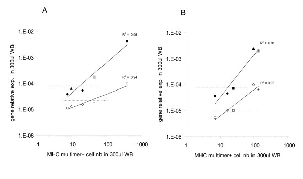
Results: Spiking experiments demonstrated that this technique could detect antigen specific cytokine gene expression from 50 cytotoxic T lymphocytes (CTL) diluted in 300 microl WB. Furthermore, the high sensitivity of this method could be confirmed ex-vivo by the successful detection of CD8+ T cell responses against HCMV, EBV and influenza virus derived HLA-A0201 restricted epitopes, which was significantly correlated with specific multimer staining. Importantly, a highly significant (p = 0.000009) correlation between hepatitis B surface antigen (HBsAg) stimulated IL-2 gene expression, as detectable in WB, and specific antibody titers was observed in donors vaccinated against hepatitis B virus (HBV) between six months and twenty years before the tests. To identify additional markers of potential clinical relevance, expression of chemokine genes was also evaluated. Indeed, HBsAg stimulated expression of MIP-1beta (CCL4) gene was highly significantly (p = 0.0006) correlated with specific antibody titers. Moreover, a longitudinal study on response to influenza vaccine demonstrated a significant increase of antigen specific IFN-gamma gene expression two weeks after immunization, declining thereafter, whereas increased IL-2 gene expression was still detectable four months after vaccination.
Conclusion: This method, easily amenable to automation, might qualify as technology of choice for high throughput screening of immune responses to large panels of antigens from cohorts of donors. Although analysis of cytokine gene expression requires adequate laboratory infrastructure, initial antigen stimulation and storage of test probes can be performed with minimal equipment and time requirements. This might prove important in "field" studies with difficult access to laboratory facilities.
Figures


Similar articles
-
[Cellular immune responses of recombinant hepatitis B (rHB) vaccine and HBsAG derived from Hansenula polymorpha cells].Zhonghua Liu Xing Bing Xue Za Zhi. 2008 Jul;29(7):706-11. Zhonghua Liu Xing Bing Xue Za Zhi. 2008. PMID: 19031766 Chinese.
-
Real time PCR for the assessment of CD8+ T cellular immune response after prophylactic vaccinia vaccination.J Clin Virol. 2007 Sep;40(1):80-3. doi: 10.1016/j.jcv.2007.04.022. Epub 2007 Jul 20. J Clin Virol. 2007. PMID: 17644471
-
Vaccination against hepatitis B in liver transplant recipients: pilot analysis of cellular immune response shows evidence of HBsAg-specific regulatory T cells.Liver Transpl. 2007 Mar;13(3):434-42. doi: 10.1002/lt.21061. Liver Transpl. 2007. PMID: 17318860 Clinical Trial.
-
Contribution of viral recombinants to the study of the immune response against the Epstein-Barr virus.Semin Cancer Biol. 2008 Dec;18(6):409-15. doi: 10.1016/j.semcancer.2008.09.001. Epub 2008 Sep 30. Semin Cancer Biol. 2008. PMID: 18938248 Review.
-
[Evolution of IGRA researches].Kekkaku. 2008 Sep;83(9):641-52. Kekkaku. 2008. PMID: 18979999 Review. Japanese.
Cited by
-
Antigen Recognition and Immune Response to Acute and Chronic Hepatitis B Virus Infection.J Inflamm Res. 2023 May 18;16:2159-2166. doi: 10.2147/JIR.S411492. eCollection 2023. J Inflamm Res. 2023. PMID: 37223107 Free PMC article. Review.
-
Considerations for accurate gene expression measurement by reverse transcription quantitative PCR when analysing clinical samples.Anal Bioanal Chem. 2014 Oct;406(26):6471-83. doi: 10.1007/s00216-014-7857-x. Epub 2014 May 25. Anal Bioanal Chem. 2014. PMID: 24858468 Free PMC article. Review.
-
Development of a one-step probe based molecular assay for rapid immunodiagnosis of infection with M. tuberculosis using dried blood spots.PLoS One. 2014 Sep 3;9(9):e105628. doi: 10.1371/journal.pone.0105628. eCollection 2014. PLoS One. 2014. PMID: 25184553 Free PMC article.
-
A whole blood monokine-based reporter assay provides a sensitive and robust measurement of the antigen-specific T cell response.J Transl Med. 2011 Aug 26;9:143. doi: 10.1186/1479-5876-9-143. J Transl Med. 2011. PMID: 21871084 Free PMC article.
-
Decoding the Immune Response and Its Biomarker B2M for High Altitude Pulmonary Edema in Rat: Implications for Diagnosis and Prognosis.J Inflamm Res. 2024 Oct 11;17:7195-7217. doi: 10.2147/JIR.S477633. eCollection 2024. J Inflamm Res. 2024. PMID: 39411751 Free PMC article.
References
-
- Harari A, Zimmerli SC, Pantaleo G. Cytomegalovirus (CMV)-specific cellular immune responses. Hum Immunol. 2004;65:500–506. - PubMed
-
- Hernandez-Fuentes MP, Warrens AN, Lechler RI. Immunologic monitoring. Immunol Rev. 2003;196:247–264. - PubMed
-
- Keilholz U, Martus P, Scheibenbogen C. Immune monitoring of T-cell responses in cancer vaccine development. Clin Cancer Res. 2006;12:2346s–2352s. - PubMed
-
- Janetzki S, Cox JH, Oden N, Ferrari G. Standardization and validation issues of the ELISPOT assay. Methods Mol Biol. 2005;302:51–86. - PubMed
Publication types
MeSH terms
Substances
LinkOut - more resources
Full Text Sources
Other Literature Sources
Research Materials
