. 2008 Oct 16:6:12.
doi: 10.1186/1476-7961-6-12.

Differential response of human basophil activation markers: a multi-parameter flow cytometry approach

Affiliations

Differential response of human basophil activation markers: a multi-parameter flow cytometry approach

Salvatore Chirumbolo et al. Clin Mol Allergy. .

Abstract

Background: Basophils are circulating cells involved in hypersensitivity reactions and allergy but many aspects of their activation, including the sensitivity to external triggering factors and the molecular aspects of cell responses, are still to be focused. In this context, polychromatic flow cytometry (PFC) is a proper tool to investigate basophil function, as it allows to distinguish the expression of several membrane markers upon activation in multiple experimental conditions.

Methods: Cell suspensions were prepared from leukocyte buffy coat of K2-EDTA anticoagulated blood specimens; about 1500-2500 cellular events for each tested sample, gated in the lymphocyte CD45dim area and then electronically purified as HLADRnon expressing/CD123bright, were identified as basophilic cells. Basophil activation with fMLP, anti-IgE and calcium ionophore A23187 was evaluated by studying up-regulation of the indicated membrane markers with a two-laser six-color PFC protocol.

Results: Following stimulation, CD63, CD13, CD45 and the ectoenzyme CD203c up-regulated their membrane expression, while CD69 did not; CD63 expression occurred immediately (within 60 sec) but only in a minority of basophils, even at optimal agonist doses (in 33% and 14% of basophils, following fMLP and anti-IgE stimulation respectively). CD203c up-regulation occurred in the whole basophil population, even in CD63non expressing cells. Dose-dependence curves revealed CD203c as a more sensitive marker than CD63, in response to fMLP but not in response to anti-IgE and to calcium ionophore.

Conclusion: Use of polychromatic flow cytometry allowed efficient basophil electronic purification and identification of different behaviors of the major activation markers. The simultaneous use of two markers of activation and careful choice of activator are essential steps for reliable assessment of human basophil functions.


Figures

Figure 1
Figure 1
Human basophils gating with the BD-FACScanto flow cytometer. Leukocytes are immunologically gated in the CD45dim-lymphocyte area and electronically captured as cellular events in HLADRnon expressing/CD123bright biparametric logarithmic plot to distinguish them from other events and identified as basophils from monocytes (Q1), plasmacytoid dendritic cells (Q2) and lymphocytes (Q3). (A) Resting cells, (B) activation with 1 μg/ml goat anti-human IgE.
Figure 2
Figure 2
Dot plots and histograms of CD63 and CD203c expression in response to fMLP. Basophils were incubated in the absence (A,B,E,G) and in the presence (C,D,F,H) of 10-7 M fMLP at 37°C for 30 minutes and gated basophils were plotted both as dot plots (A,B,C,D) and as histograms (E,F,G,H).
Figure 3
Figure 3
Dot plots and histograms of CD63 and CD203c expression in response to anti-IgE. Basophils were incubated in the absence (A,B,E,G) and in the presence (C,D,F,H) of 10 μg/ml goat anti-IgE at 37°C for 30 minutes and gated basophils were plotted both as dot plots (A,B,C,D) and as histograms (E,F,G,H).
Figure 4
Figure 4
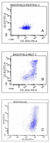
Biparametric dot plots of CD63 and CD203c expression. Biparametric dot plots showing CD63 and CD203c in resting basophils (A) and after activation with 100 nM fMLP (B) or with 10 μg/ml goat anti-IgE (C).
Figure 5
Figure 5
Intracellular CD63. Dot plots (A,C) and histograms (B,D) of CD63-associated fluorescence in normal (A,B) and saponin-permeabilized human basophils (C,D). See text for methods.
Figure 6
Figure 6
fMLP and anti-IgE dose response of CD63 and CD203c expression. fMLP (a) and goat anti-human IgE dose response (b) of the activation markers CD63 and CD203c in a typical triplicate experiment of four performed. Basophils were incubated for 30 minutes at 37°C.
Figure 7
Figure 7
Time course of CD63 and CD203c expression. Basophils were incubated with 10-7 M fMLP at 37°C and activation stopped with cold HEPES-buffered solution containing 2.8 mM Na2-EDTA.
Figure 8
Figure 8
Expression of CD203c in CD63non expressing human basophils. Basophils were incubated for 30 minutes at 37°C in the presence of different concentrations of fMLP and the MFI of CD203c was evaluated in the subset of CD63non expressing basophils.
Figure 9
Figure 9
Dose response curves of CD63 and CD203c following stimulation with A23187. Basophils were incubated for 30 minutes at 37°C in the presence of increasing doses of the calcium ionophore A23187.

References

    1. Boumiza R, Debard AL, Monneret G. The basophil activation test by flow cytometry: recent developments in clinical studies, standardization and emerging perspectives. Clin Mol Allergy. 2005;3:9. doi: 10.1186/1476-7961-3-9. - DOI - PMC - PubMed
    1. Triggiani M, Marone G. Basophil's secrets revealed by flow cytometry. Allergy. 2006;61:1025–1027. doi: 10.1111/j.1398-9995.2006.01217.x. - DOI - PubMed
    1. Ebo DG, Sainte-Laudy J, Bridts CH, Mertens CH, Hagendorens MM, Schuerwegh AJ, De Clerck LS, Stevens WJ. Flow-assisted allergy diagnosis: current applications and future perspectives. Allergy. 2006;61:1028–1039. doi: 10.1111/j.1398-9995.2006.01039.x. - DOI - PubMed
    1. Abuaf N, Rostane H, Rajoely B, Gaouar H, Autegarden JE, Leynadier F, Girot R. Comparison of two basophil activation markers CD63 and CD203c in the diagnosis of amoxicillin allergy. Clin Exp All. 2008;38:921–928. doi: 10.1111/j.1365-2222.2008.02960.x. - DOI - PubMed
    1. de Week AL, Sanz ML, Gamboa PM, Aberer W, Bienvenu J, Bianca M, Demoly P, Ebo DG, Mayorga L, Monneret G, Sainte-Laudy J. Diagnostic tests based on human basophils: more potentials and perspectives than pitfalls: II Technical issues. J Investig Allergol Clin Immunol. 2008;18:143–155. - PubMed