. 2008 Oct 7;52(15):1222-30.
doi: 10.1016/j.jacc.2008.07.012.

Repolarization and activation restitution near human pulmonary veins and atrial fibrillation initiation: a mechanism for the initiation of atrial fibrillation by premature beats

Affiliations

Repolarization and activation restitution near human pulmonary veins and atrial fibrillation initiation: a mechanism for the initiation of atrial fibrillation by premature beats

Sanjiv M Narayan et al. J Am Coll Cardiol. .

Abstract

Objectives: The authors sought to study mechanisms to explain why single premature atrial complexes (PACs) from the pulmonary veins (PVs) may initiate human atrial fibrillation (AF).

Background: Theoretically, single PACs may initiate AF if the rate response of action potential duration (APD) restitution has a slope >1. However, human left atrial APD restitution and its relationship to AF have not been studied. We hypothesized that an APD restitution slope >1 near PVs explains the initiation of clinical AF.

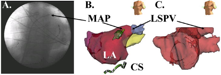
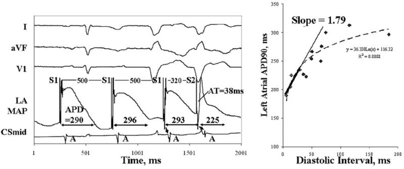
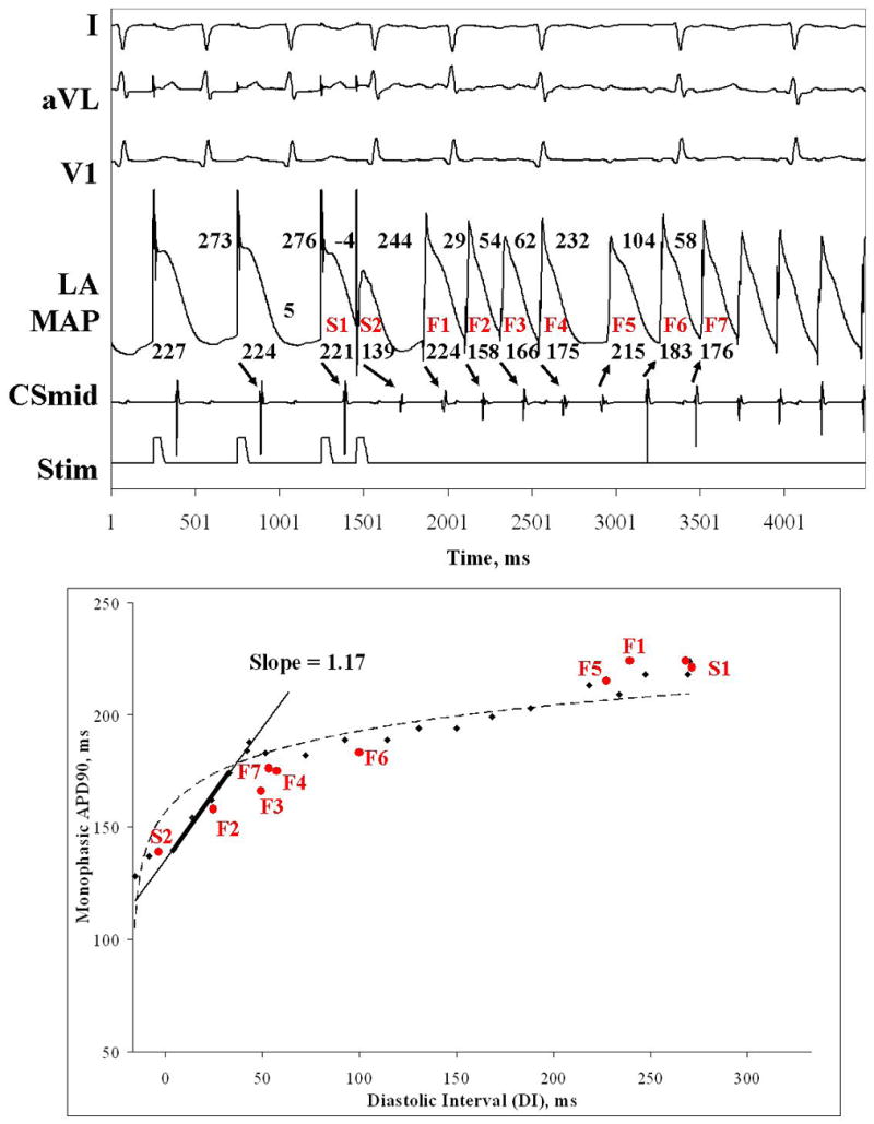
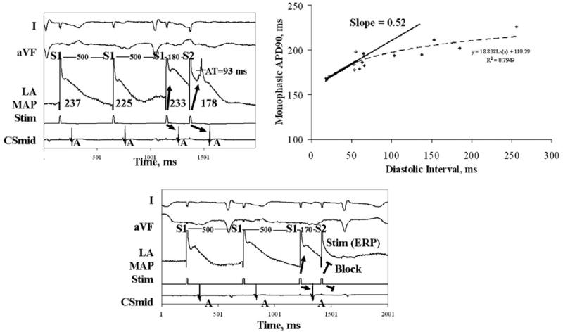
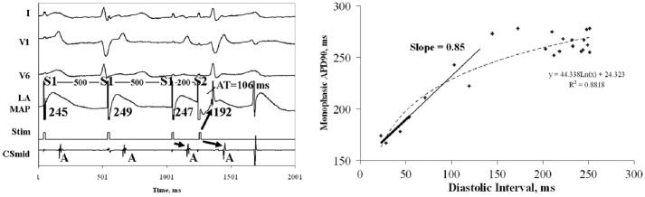
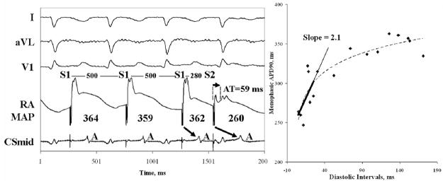
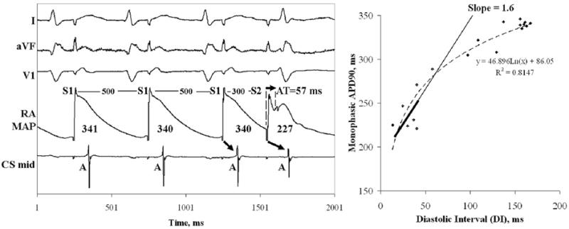
Methods: We studied 27 patients with paroxysmal and persistent (n = 13) AF. We advanced monophasic action potential catheters transseptally to superior PVs. Restitution was plotted as APD of progressively early PACs against their diastolic interval (DI) from prior beats. Activation time restitution was measured using the time from the pacing artifact to each PAC.

Results: Compared with paroxysmal AF, patients with persistent AF had shorter left atrial APD and effective refractory period (p = 0.01). In paroxysmal AF, maximum left atrial APD restitution slope was 1.5 +/- 0.4; and 12 of 13 patients had slope >1 (p < 0.001). In persistent AF, PACs encountered prolonged activation for a wider range of beats than in paroxysmal AF (p = 0.01), which prolonged DI and flattened APD restitution (slope 0.7 +/- 0.2; p < 0.001); no patient had APD restitution slope >1. A single PAC produced AF in 5 patients; in all, an APD restitution slope >1 caused extreme APD oscillations after the PAC, then AF.

Conclusions: In patients with paroxysmal AF, maximum APD restitution slope >1 near the PVs enables single PACs to initiate AF. However, patients with persistent AF show marked dynamic activation delay near PVs that flattens APD restitution. Studies should determine how regional APD and conduction dynamics contribute to the substrates of persistent AF.


Figures

Figure 1
Figure 1
Left Atrial Monophasic Action Potential (MAP) recording site, in a 74 year old man with persistent AF and LA diameter 48 mm. (A) Fluoroscopy (Left Anterior Oblique 30o) showing MAP catheter within a trans-septal sheath near the left superior PV (LSPV), and the coronary sinus (CS) catheter; (B) Digital Reconstruction of LA confirming MAP catheter in the LSPV antrum (NavX, St Jude Medical, CA). (C) Segmented 64-slice computed tomogram imported into NavX for positional reference.
Figure 2
Figure 2
Left Atrial Action Potentials During Constant Pacing (S1) and PACs (S2) in patients with (A) Paroxysmal AF; and (B) Persistent AF. For each PAC, stimulus artifact (“Stim”) and phases 1, 2 and 3 of the AP are labeled. Activation time (AT) spans the time from the stimulus artifact to the computed dV/dt maximum of phase 0 (not labeled), and is 25 ms for A and 50 ms in B. Bipolar atrial electrograms are labeled “A” in the coronary sinus (CS). (C) APD90 measurement, calculated as 90% repolarization from phase II voltage to the baseline.
Figure 3
Figure 3. Steep Left Atrial APD Restitution in Paroxysmal AF
This PAC is delivered just outside the ERP (500/320 ms) and results in DI 6 ms and APD restitution slope = 1.8 (same patient as figure 2A). The thickened line represents the DI range for which slope was calculated.
Figure 4
Figure 4. PAC Induces Paroxysmal AF In a Patient with Steep APD Restitution
(A) Very early PAC (DI -4 ms) followed by a pause then AF. (B) Steep Restitution May Explain PAC-Induced AF (in red). S1, S2, F1 and F2 show marked APD oscillations, because of steep APD restitution (slope = 1.2; slope > 3 by monoexponential fit), then AF onset. AF cycles continue to track restitution, even though wavelets meander (altered activation sequence after F4).
Figure 5
Figure 5. Left Atrial APD Restitution Is Less Steep in Persistent AF
(A) This PAC is early (coupling interval 180 ms), yet significant AT delay (93 ms) enables capture (see blocked stimulus artifact at ERP, 170 ms). This prolongs DI and flattens APD restitution. (B) This early PAC also encounters AT delay (106ms). APD restitution has maximum slope 0.85 (i.e. also not steep; same patient as fig 2B). In both patients, FRP (minimum interval from prior beat to PAC) is longer than ERP (minimum distance between stimuli). See table II.
Figure 6
Figure 6. Right Atrial APD restitution has slope > 1 Without AT Prolongation
In (A) Paroxysmal AF, PACs and APD restitution for a 74 year old man with LA diameter 45 mm and AF for 3 years; (B) Persistent AF, in the same patient as figure 5B. Compared to left atrial data in this patient, ERP (280 ms vs 200 ms) and APD90 (346 ms vs 275 ms) were longer, and AT delay (57 ms vs 106 ms) and minimum DI (13 vs 23 ms) were shorter. See table II.
Figure 7
Figure 7. Left Atrial Activation Prolongs for a Wider Range of Beats in Persistent than Paroxysmal AF
In (A) Paroxysmal AF, AT prolongs only at very early DI (<20 ms; i.e. preserved conduction restitution), seen in actual and normalized plots. (B) Persistent AF, AT prolongs at longer DI (< 108 ms; i.e. broad conduction restitution) in both plots. See table III.

References

    1. Haissaguerre M, Jais P, Shah DC, et al. Spontaneous Initiation of Atrial Fibrillation by Ectopic Beats Originating in the Pulmonary Veins. N Engl J Med. 1998;339:659–666. - PubMed
    1. Haissaguerre M, Sanders P, Hocini M, et al. Changes in Atrial Fibrillation Cycle Length and Inducibility During Catheter Ablation and Their Relation to Outcome. Circulation. 2004;109:3007–3013. - PubMed
    1. Gerstenfeld EP, Sauer W, Callans DJ, et al. Predictors of success after selective pulmonary vein isolation of arrhythmogenic pulmonary veins for treatment of atrial fibrillation. Heart Rhythm. 2006;3:165–170. - PubMed
    1. Haissaguerre M, Sanders P, Hocini M, et al. Catheter Ablation of Long-Lasting Persistent Atrial Fibrillation: Critical Structures for Termination. Journal of Cardiovascular Electrophysiology. 2005a;16:1125–1137. - PubMed
    1. Gong Y, Xie F, Stein K, et al. Mechanism underlying initiation of paroxysmal atrial flutter/atrial fibrillation by ectopic foci: a simulation study. Circulation. 2007;115:2094–102. - PubMed

Publication types