Review
. 2008 Nov;16(4):697-710, ix-x.
doi: 10.1016/j.mric.2008.07.005.

Advances in MR spectroscopy of the prostate

Affiliations
Review

Advances in MR spectroscopy of the prostate

John Kurhanewicz et al. Magn Reson Imaging Clin N Am. 2008 Nov.

Abstract

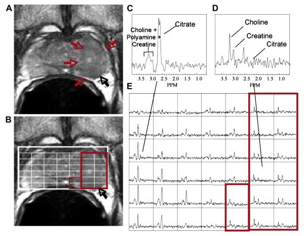
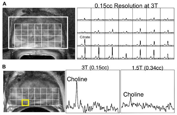
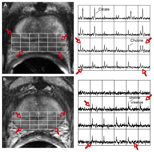
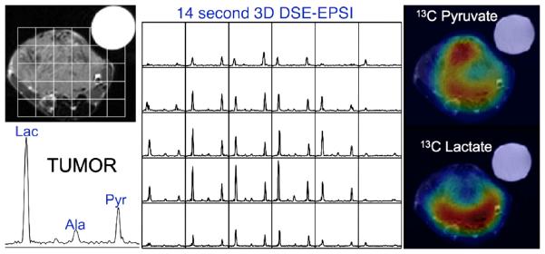
Commercial MR imaging/magnetic resonance spectroscopic imaging (MRSI) packages for staging prostate cancer on 1.5-T MR scanners are now available. The technology is becoming mature enough to begin assessing its clinical utility in selecting, planning, and following prostate cancer therapy. Before therapy, 1.5-T MR imaging/MRSI has the potential to improve the local evaluation of prostate cancer presence and volume and has a significant incremental benefit in the prediction of pathologic stage when added to clinical nomograms. After therapy, two metabolic biomarkers of effective and ineffective therapy have been identified and are being validated with 10-year outcomes. Accuracy can be improved by performing MR imaging/MRSI at higher magnetic field strengths, using more sensitive hyperpolarized (13)C MRSI techniques and through the addition of other functional MR techniques.


Figures

Figure 1
Figure 1
(a) A) A reception-profile corrected T2 weighted FSE axial image taken from a volume MRI/MRSI data set demonstrating a large region of hypointensity in the left midgland (red arrows) with suspected extracapsular extension (black arrow). The selected volume for spectroscopy (bold white box) and a portion of the 16 × 8 × 8 spectral phase encode grid overlaid (fine white line) on the T2 weighted image (B) with the corresponding 0.3 cm3 proton spectral array (E). Spectra in regions of cancer (D, red box) demonstrate dramatically elevated choline, an absence of citrate and polyamines relative to regions of healthy peripheral zone tissue (C). In this fashion, metabolic abnormalities can be correlated with anatomic abnormalities from throughout the prostate. The strength of the combined MRI/MRSI exam has been found to be when changes in all three metabolic markers (choline, polyamines and citrate) and imaging findings are concordant for cancer.
Figure 2
Figure 2
Prostate MRI/MRSI data obtained on a 3 Tesla scanner with the j-refocused, MLEV-PRESS (Point RESolved Spectroscopy) MRSI sequence (101). A) T2-weighted MRI and a 3D MR spectral array are shown for a pre-therapy patient. The 3D MRSI data were acquired in 17 minutes with a spatial resolution of 0.15 cm3 using the specialized acquisition sequence to obtain upright citrate resonances at an echo time of 85ms on a 3 Tesla MR scanner. B) 3T MRI and MRSI data are shown for the same prostate cancer patient with biopsy proven cancer in the right midgland. The higher resolution obtainable at 3T MRSI (middle) depicted more clearly the elevated choline levels as compared to the corresponding 1.5T data (right) acquired on the same day.
Figure 3
Figure 3
(A) Endorectal coil Fast Spin Echo T2 weighted image from the midgland of prostate demonstrating bilateral low signal intensity (red arrows) in the regions of biopsy proven prostate cancer. The overlying white grid shows the locations where the MRSI spectra are taken from (right). Regions of prostate cancer (red arrow) demonstrate elevated choline and reduced citrate relative to regions of healthy tissue. (B) A T2 weighted image and corresponding spectral array taken from the same location as in (A) four years after combined radiation and androgen deprivation therapy. There are a number of voxels demonstrating metabolic atrophy consistent with effective treatment; however, there remain bilateral regions of elevated choline that was confirmed to be recurrent prostate cancer at biopsy.
Figure 4
Figure 4
This study of a TRAMP prostate model tumor using the HyperSense system for pre-polarization demonstrated not only high 13C lactate in the tumor but also differences in metabolite distributions in the tumor. The data were acquired in 14 seconds and show high uptake of hyperpolarized pyruvate throughout much of the tumor, except for a presumably necrotic region at the center. The lactate image demonstrated a focus of high metabolic activity in the posterior aspect of the tumor, which is indicative of biologically aggressive cancer.

References

    1. Hricak H, White S, Vigneron D, et al. Carcinoma of the prostate gland: MR imaging with pelvic phased-array coils versus integrated endorectal--pelvic phased-array coils. Radiology. 1994;193:703–709. - PubMed
    1. Kurhanewicz J, Vigneron DB, Hricak H, Narayan P, Carroll P, Nelson SJ. Three-dimensional H-1 MR spectroscopic imaging of the in situ human prostate with high (0.24-0.7-cm3) spatial resolution. Radiology. 1996;198:795–805. - PubMed
    1. Tuan-Khanh CT, Vigneron DB, Sailasuta N, et al. Reducing Chemical Shift Errors and Conforming PRESS-CSI Selection with Very Selective Saturation (VSS) Pulses. JMRI. 1999
    1. Schricker AA, Pauly JM, Kurhanewicz J, Swanson MG, Vigneron DB. Dualband spectral-spatial RF pulses for prostate MR spectroscopic imaging. Magn Reson Med. 2001;46:1079–1087. - PubMed
    1. Cunningham CH, Vigneron DB, Chen AP, et al. Design of symmetric-sweep spectral-spatial RF pulses for spectral editing. Magn Reson Med. 2004;52:147–153. - PubMed

Publication types