Review
. 2008 Dec;33(12):609-20.
doi: 10.1016/j.tibs.2008.09.003. Epub 2008 Oct 14.

Rising from the RecQ-age: the role of human RecQ helicases in genome maintenance

Affiliations
Review

Rising from the RecQ-age: the role of human RecQ helicases in genome maintenance

Vilhelm A Bohr. Trends Biochem Sci. 2008 Dec.

Abstract

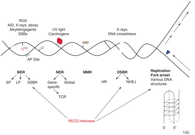
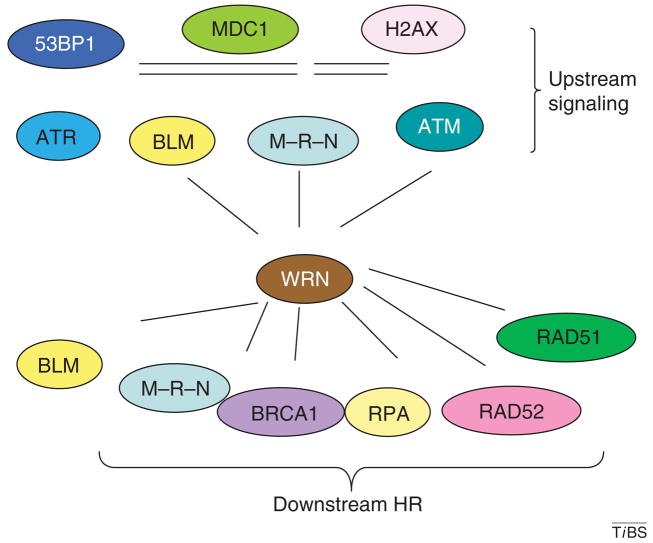
The RecQ helicases are guardians of the genome. Members of this conserved family of proteins have a key role in protecting and stabilizing the genome against deleterious changes. Deficiencies in RecQ helicases can lead to high levels of genomic instability and, in humans, to premature aging and increased susceptibility to cancer. Their diverse roles in DNA metabolism, which include a role in telomere maintenance, reflect interactions with multiple cellular proteins, some of which are multifunctional and also have very diverse functions. The results of in vitro cellular and biochemical studies have been complimented by recent in vivo studies using genetically modified mouse strains. Together, these approaches are helping to unravel the mechanism(s) of action and biological functions of the RecQ helicases.


Figures

Figure 1
Figure 1
The RecQ helicase family. Schematic diagram of RecQ helicases from diverse species. Conserved domains are aligned and color-coded, as indicated at the bottom of the figure. The size of each protein species in amino acids is indicated on the right. Protein name and species of origin is indicated on the left. The five human RecQ helicases are indicated by the red box. RECQ5 exists in three isoforms, the β form is shown.
Figure 2
Figure 2
RecQ enzymatic functions and possible functional roles. (a) Schematic summary of the enzymatic properties of WRN and BLM. (b) Preferred DNA substrates of WRN and BLM are shown. (c) Proposed functional roles of WRN and BLM at a stalled DNA replication fork. Possible outcomes are indicated. DNA damage is present at the replication fork.
Figure 3
Figure 3
WRN and BLM functional protein interactions. (a) Protein interacting partners of WRN are indicated according to functional effect. Note that this list only includes functional protein interactions (i.e. interactions that modulate the function of one or both of the protein partners (see elsewhere for further discussion). The target WRN activity is shown in parentheses. (i) Proteins that stimulate WRN. (ii) Proteins that are stimulated by WRN. (iii) Proteins that inhibit WRN. (iv) Proteins that are inhibited by WRN. Proteins that have analogous interactions with both WRN and BLM are shown in red. (b) Same as part (a), except for BLM. Abbreviations: exo, exonuclease; FLAP, DNA flap; hel, helicase; TOP1, topoisomerase I.
Figure 4
Figure 4
Clinical features of BS and WS.
Figure 5
Figure 5
Eukaryotic DNA repair. Schematic diagram summarizing eukaryotic DNA-repair pathways and their target DNA lesions. Abbreviations: AID, activation-induced cytidine deaminase; AP, apurinic or apyrimidinic; Gox, 8-oxoG; MM, mismatch; NHEJ, non-homologous end-joining; SSBR, single-strand break repair; TCR, transcription-coupled repair; U, uracil. See text for discussion of RecQ helicase roles in DNA repair.
Figure 6
Figure 6
WRN in the DSBR DNA-damage response. WRN protein interactions indicate possible roles in early and late steps of the response to replication stress. Several of the proteins involved in the formation of the DNA double-strand breaks repair formation are shown under upstream signaling, and several of the proteins involved in the DNA-repair process of homologous recombination are shown. Abbreviations: 53BP1, 53 binding protein; MDC1, mediator of DNA damage checkpoint 1; M–R–N, MRE11–RAD50–NBS1 complex.
Figure 7
Figure 7
WRN post-translational modification. Schematic diagram summarizing post-translational modifications of WRN including phosphoryation on Ser/Thr or Tyr (upper half of figure), sumoylation (bottom left), acetylation (bottom middle) and oxidation (bottom right). Probable mediators and modifiers of WRN post-translational modifications are indicated. Functional effects of post-translational modifications are indicated, where known. The ‘?’ indicates uncertainty. Arrows up or down: two arrows indicate stronger reaction than one arrow.

References

    1. German J. Bloom’s syndrome. XX The first 100 cancers. Cancer Genet Cytogenet. 1997;93:100–106. - PubMed
    1. Hickson ID. RecQ helicases: caretakers of the genome. Nat Rev Cancer. 2003;3:169–178. - PubMed
    1. Wu L, Hickson ID. DNA helicases required for homologous recombination and repair of damaged replication forks. Annu Rev Genet. 2006;40:279–306. - PubMed
    1. Mohaghegh P, et al. The Bloom’s and Werner’s syndrome proteins are DNA structure-specific helicases. Nucleic Acids Res. 2001;29:2843–2849. - PMC - PubMed
    1. von Kobbe C, et al. Colocalization, physical, and functional interaction between Werner and Bloom syndrome proteins. J Biol Chem. 2002;277:22035–22044. - PubMed

Publication types

MeSH terms

LinkOut - more resources