Review
. 2009 Feb;87(3):133-70.
doi: 10.1016/j.pneurobio.2008.09.009. Epub 2008 Sep 24.

Nanotechnology, nanotoxicology, and neuroscience

Affiliations
Review

Nanotechnology, nanotoxicology, and neuroscience

Won Hyuk Suh et al. Prog Neurobiol. 2009 Feb.

Abstract

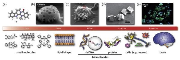
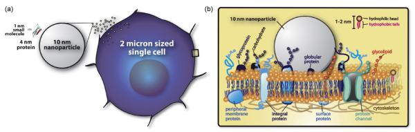
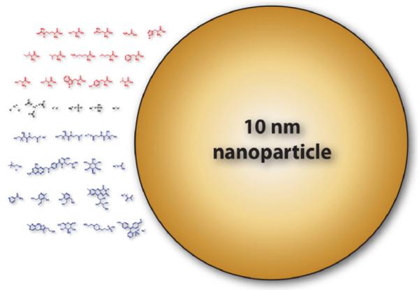
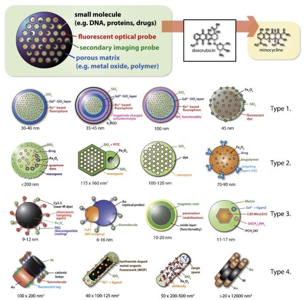
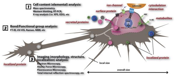
Nanotechnology, which deals with features as small as a 1 billionth of a meter, began to enter into mainstream physical sciences and engineering some 20 years ago. Recent applications of nanoscience include the use of nanoscale materials in electronics, catalysis, and biomedical research. Among these applications, strong interest has been shown to biological processes such as blood coagulation control and multimodal bioimaging, which has brought about a new and exciting research field called nanobiotechnology. Biotechnology, which itself also dates back approximately 30 years, involves the manipulation of macroscopic biological systems such as cells and mice in order to understand why and how molecular level mechanisms affect specific biological functions, e.g., the role of APP (amyloid precursor protein) in Alzheimer's disease (AD). This review aims (1) to introduce key concepts and materials from nanotechnology to a non-physical sciences community; (2) to introduce several state-of-the-art examples of current nanotechnology that were either constructed for use in biological systems or that can, in time, be utilized for biomedical research; (3) to provide recent excerpts in nanotoxicology and multifunctional nanoparticle systems (MFNPSs); and (4) to propose areas in neuroscience that may benefit from research at the interface of neurobiologically important systems and nanostructured materials.


Figures

Fig. 1
Fig. 1
The sizes of biologically relevant entities. (Top row above scale bar) From left to right: (a) Potent Alzheimer’s disease candidate drug, dehydroevodiamine HCl (DHED) X-ray crystal structure, (b and c) porous metal oxide microspheres being endocytosed by BV2 microglia cell (close-up and low magnification) SEM images, (d and e) SEM and fluorescence micrograph of DHED microcrystals (DHED is blue-green luminscent). (Bottom row below the scale bar) Left to right: Small molecules, such as dopamine, minocycline, mefenamic acid, DHED, and heme, are ∼1 nm or smaller. The lipid bilayer is a few nanometers thick. A biomolecule such as a (22 bp) microRNA and a protein is only a few nanometers in size. A single cell or neuron is tens or hundreds of microns in size. Illustration of a human brain which is tens of centimeters in size.
Fig. 2
Fig. 2
Size matters. (a) Compared to a 10 nm nanoparticle, proteins (e.g. APP; X-ray crystal structure obtained from www.pdb.org (Berman et al., 2000), protein ID 2FKL; visualization done by Accelrys Discovery Studio Visualization 1.7 software) and small molecules (e.g. DHED) are small in size and volume. A mammalian cell which is made up of proteins, nucleic acids, and other small to large molecules is thousand times larger in volume and size compared to a 10 nm nanoparticle. (b) Cell membrane incorporating various proteins and a single 10 nm nanoparticle.
Fig. 3
Fig. 3
Published papers in nanomaterials synthesis papers published in 1970–2007. Number of publications was obtained from ISI Web of Science (one of Thomson Scientific databases and part of Web of Knowledge) using a combination of search terms that represent nanomaterial and synthesis.
Fig. 4
Fig. 4
Contents of DMEM vs. 10 nm nanoparticle. Red chemical structures (first three rows) represent amino acids, black chemical structures (fourth row) represent inorganic salts, and blue structures (rows 5–8) represent vitamins and other small organic molecules. The contents information of DMEM (Dulbecco’s Modified Eagle’s Medium) were readily available on-line at various biochemical vendor websites such as HyClone and Sigma—Aldrich.
Fig. 5
Fig. 5
Cell and particle interactions. Toxicological effects of nanomaterials can be simplified into eight events as shown in the illustration above but limiting the interaction between a nanoparticle and a cell to eight events is an over simplification and the details of actual phenomena that are happening at the interfaces are very difficult to analyze and understand. (1) Reactive oxygen species products such as superoxide (O2-) and hydroxyl radical (OH) whether it is inside or outside can be key factors in nanostructured materials toxicological effects (Nel et al., 2006). Cell membrane integrity leading to cell survivability will be affected by ROS produced by a nanoparticle smaller than a cell (red particle) as shown. (2) Event 2 represents the situation where a nanoparticle is internalized and then creates ROS products (Nel et al., 2006). (3) Particle dissolution affecting cellular function after nanoparticle internalization is event 3 (Borm et al., 2006). (4) Event 4 represents any mechanical damage to sub-cellular units such as the lysosome, endoplasmic reticulum, and nucleus (Yamamoto et al., 2004). (5) Different functional groups and surface electronic structures arising from different nanostructured materials will determine the level of interaction between the nanoparticles and their surroundings which is represented by event 5 (Karakoti et al., 2006; Kostarelos et al., 2007). (6) Overall size of the particle can play an important role as represented by event 6 since large particles can potentially induce permanent damage to the cell membrane while small particles can pass through the membrane and do harm inside cell (Yoshida et al., 2003). (7) Non-spherical particles, on the other hand, might have a different biological response compared to the spherical nanoparticles which is shown as event 7 (Geng et al., 2007). (8) Event 8 represents dissolution characteristics of the nanomaterials outside the cell which can affect the cell in various ways (event 8) (Borm et al., 2006).
Fig. 6
Fig. 6
Nanobiocomposite formed from a nanoparticle (sub-micron) and a nanobacteria (e.g. mycoplasma; sub-500 nm). This event is probable to happen under biogenic conditions where polyelectrolytes (e.g. peptide) and soluble ionic species (e.g. Ca2+, Na+) are readily available. Sub-micron engineered nanoparticles can form new composite materials with mycoplasma and the new nanobiocomposite material can have vastly different chemistries and physical properties which will lead to different biological properties.
Fig. 7
Fig. 7
Spherical and non-tubular carbon nanomaterials. Sub-100 nm carbon nanoparticles that are other than C60 or carbon nanotubes will offer another set of tools for neuroscientist as well as other biologists. Illustrations were prepared based on data, schemes, and figures appearing in the references with permission from the publisher.
Fig. 8
Fig. 8
Controlling cell function by microscale patterns and nanowires. Details are provided for the top three illustrations in the maintext. Making sub-micron patterns as well as functionalizing the sub-patterns with unique nanostructures such as wires and pores will be very interesting to utilize in neuroscience, especially studying interacting neurons and neuronal implants in vivo.
Fig. 9
Fig. 9
Multifunctional nanoparticle systems (MFNPS) for biomedical applications. MFNPSs can be divided into four distinctive types. Type 1 is non-porous but spherical SiO2 based sub-100 nm nanoparticles with two or more components. Type 2 is sub-200 nm spherical nanoparticles that is either porous or can incorporate and, in time, release small molecules such as drug molecules. Type 3 is sub-20 nm nanoparticles with functionalizable ligands or biomolecules stabilized (passivated) onto the nanoparticles and are, in most cases, first synthesized in organic conditions which offer good size control and then phase exchanged to become dispersable in aqueous media. Finally, type 4 is non-spherical nanoparticle systems that have multiple components such as fluorescent tags and antibodies. Illustrations were prepared based on data, schemes, and figures appearing in the references of Table 5 with permission.
Fig. 10
Fig. 10
Analysis of a cell. Sub-components of a cell include (but not exclusive) nucleic acids, membrane fractions, proteins (e.g. secreted, surface displaying, localized), ion channels, and cytoskeletal components. Considering the nature of such sub-cellular components and products three categories of analyses can be drawn: (1) cell content (elemental) analysis, (2) chemical bond/functional group analysis, (3) imaging (morphology, structure, localization) analysis.
Fig. 11
Fig. 11
Nanoscale imaging of biomolecules and inorganic materials. (Top row) high aspect ratio nanomaterials (e.g. fibrillar, tubular, and rod shaped); (bottom row) low aspect ratio nanostructures (e.g. oligomeric, spherical, and sub-100 nm nanoparticles). (a) AFM image of Aβ, tubular form. (b) TEM image of titanium oxide nanotubes. (c) Illustration representing crystallization schemes for high aspect ratio nanomaterials. (d) AFM image of Aβ, oligomeric form. (e) TEM image of titanium oxide nanoparticles. (f) Illustration representing crystallization schemes of spherical nanomaterials. (a) and (d) (the AFM data) were adapted from reference Heo et al. (2007) with permission from the publisher.
Fig. 12
Fig. 12
RNA interference vehicles. Various shapes and forms are used as tools to deliver RNA that will selectively silence gene translation; examples include dendrimers, copolymers, nucleic acid decorated Au NPs, nanocomposite spheres, multifunctional QDs, carbon nanotube arrays, and nanocircular RNAs. Illustrations were prepared based on data, schemes, and figures appearing in the references with permission from the publisher.

References

    1. Aaron J, Nitin N, Travis K, Kumar S, Collier T, Park SY, Jose-Yacaman M, Coghlan L, Follen M, Richards-Kortum R, Sokolov K. Plasmon resonance coupling of metal nanoparticles for molecular imaging of carcinogenesis in vivo. J. Biomed. Opt. 2007;12:034007. - PubMed
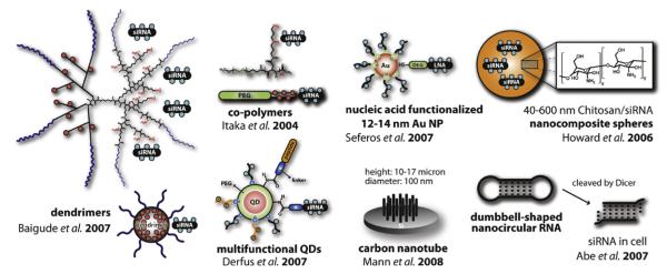
    1. Abe N, Abe H, Ito Y. Dumbbell-shaped nanocircular RNAs for RNA interference. J. Am. Chem. Soc. 2007;129:15108–15109. - PubMed
    1. Adams LK, Lyon DY, Alvarez PJJ. Comparative eco-toxicity of nanoscale TiO2, SiO2, and ZnO water suspensions. Water Res. 2006;40:3527–3532. - PubMed
    1. Afaq F, Abidi P, Matin R, Rahman Q. Activation of alveolar macrophages and peripheral red blood cells in rats exposed to fibers/particles. Toxicol. Lett. 1998a;99:175–182. - PubMed
    1. Afaq F, Abidi P, Matin R, Rahman Q. Cytotoxicity, pro-oxidant effects and antioxidant depletion in rat lung alveolar macrophages exposed to ultrafine titanium dioxide. J. Appl. Toxicol. 1998b;18:307–312. - PubMed

Publication types