

Review
doi: 10.1074/jbc.R800054200.
Epub 2008 Oct 16.
Ryanodine receptor structure: progress and challenges
Affiliations
- PMID: 18927076
- PMCID: PMC3837402
- DOI: 10.1074/jbc.R800054200
Item in Clipboard
Review
Ryanodine receptor structure: progress and challenges
J Biol Chem.
.
No abstract available
Figures
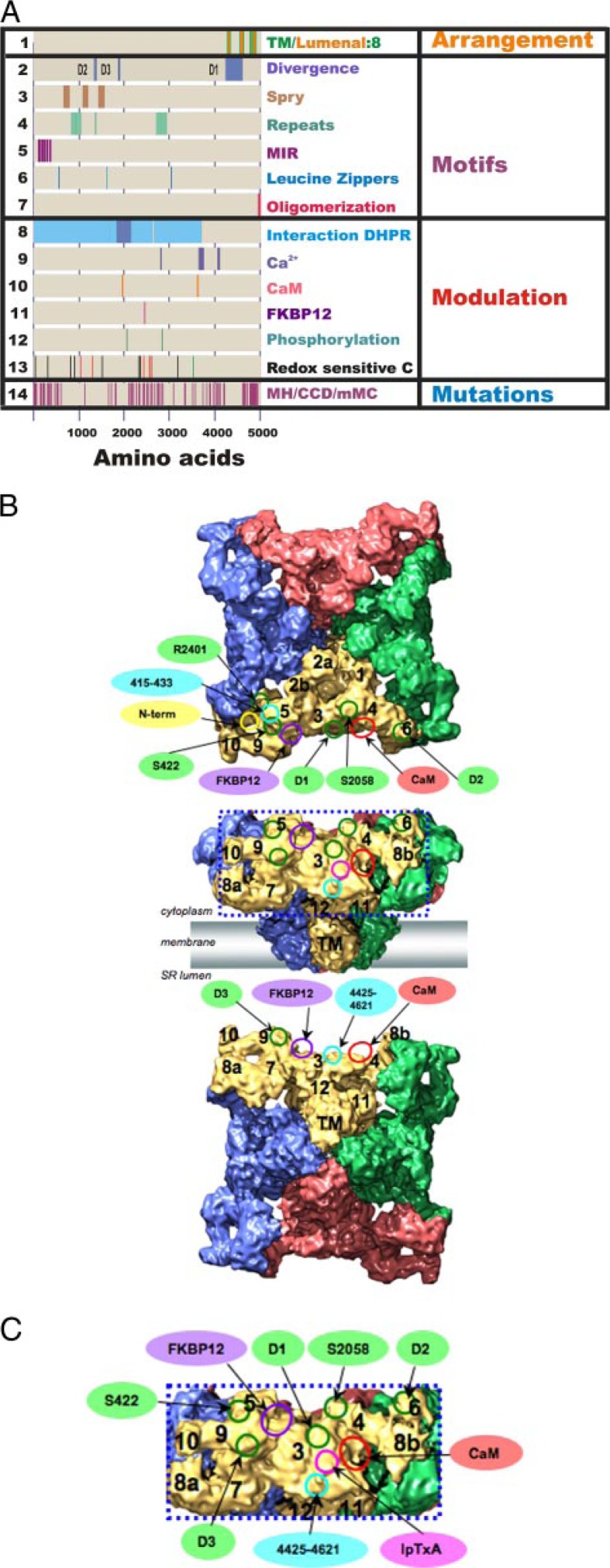
Correlating primary sequence motifs with subdomains in the 9.6-Å three-dimensional structure.
A, sites identified in the primary sequence of RyR1. DHPR, dihydropyridine receptor; MH, malignant hyperthermia; CCD, central core disease; mMC, multiminicore disease. B, cryo-EM map viewed from the cytoplasm (upper panel) and from the sarcoplasmic reticulum (SR) lumen (lower panel). RyR1 subunits are color-coded; putative subregions are indicated. Circles indicate the ligand-binding sites and RyR sequences on the three-dimensional map of RyR by single-particle cryo-EM: red, CaM-binding site; purple, FKBP12-binding site; magenta, imperatoxin A (IpTxA)-binding site; green, sequences mapped by reconstructions of expressed RyR2-GFP fusion proteins; cyan, sequence-specific antibody-binding sites; yellow, the position of the glutathione S-transferase tag fused to the N terminus of RyR3. C, enlargement of the boxed region in B. Ligand-binding sites are labeled as described for B.
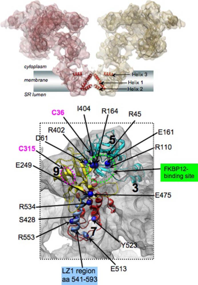
RyR1 Ca2+ release channel closed-state structure at 9.6-Å resolution (2, 4).
A, two subunits of RyR1 are shown as semitransparent surfaces. α-Helices in the Ca2+ conduction pathway of the TM region are annotated. SR, sarcoplasmic reticulum. B, malignant hyperthermia/central core disease mutations are mapped to the homology models for the N-terminal region of RyR1 (aa 12–565), localized to the clamp-shaped region of the channel structure (aa numbers correspond to Swiss-Prot accession number P11716). The FKBP12-binding region is indicated in green; the LZ1 region is shown in blue; and hyper-reactive cysteines (Cys36 and Cys315) are indicated in magenta. The location of Cys315 (magenta circle) is tentatively assigned because sequence Ala311–Glu343 was excluded from the homology model due to lack of a structural template for this region.
References
-
- Serysheva II, Chiu W, Ludtke SJ. Methods Cell Biol. 2007;79:407–435. - PubMed
Publication types
MeSH terms
Substances
Grants and funding
LinkOut - more resources
Full Text Sources
Other Literature Sources
Molecular Biology Databases
