. 2008 Nov;135(22):3707-17.
doi: 10.1242/dev.026773. Epub 2008 Oct 16.

The development of motor coordination in Drosophila embryos

Affiliations

The development of motor coordination in Drosophila embryos

Sarah Crisp et al. Development. 2008 Nov.

Abstract

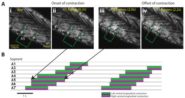
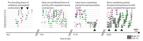
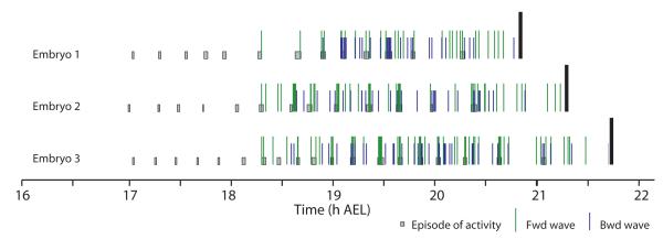
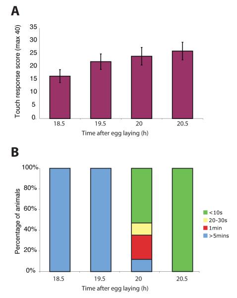
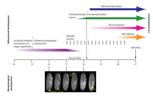
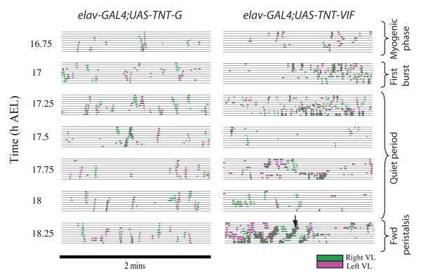
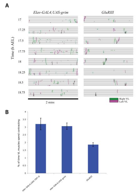
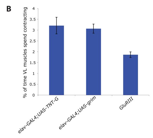
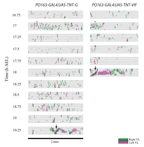
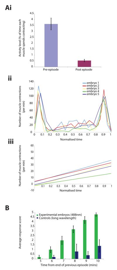
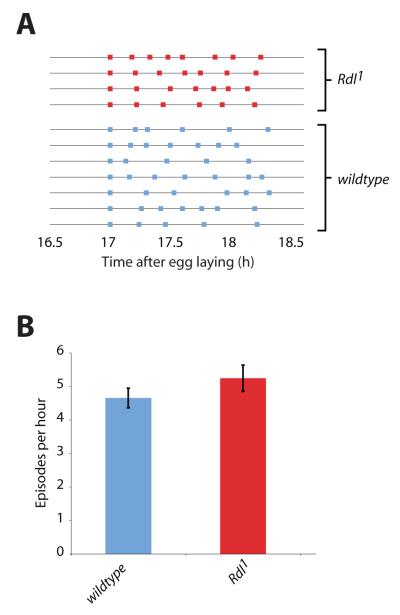
We used non-invasive muscle imaging to study the onset of motor activity and emergence of coordinated movement in Drosophila embryos. Earliest movements are myogenic, and neurally controlled muscle contractions first appear with the onset of bursting activity 17 hours after egg laying. Initial episodes of activity are poorly organised and coordinated crawling sequences only begin to appear after a further hour of bursting. Thus, network performance improves during this first period of activity. The embryo continues to exhibit bursts of crawling-like sequences until shortly before hatching, while other reflexes also mature. Bursting does not begin as a reflex response to sensory input but appears to reflect the onset of spontaneous activity in the motor network. It does not require GABA-mediated transmission, and, by using a light-activated channel to excite the network, we demonstrate activity-dependent depression that may cause burst termination.


Figures

Figure 1
Figure 1. Recording and analysing muscle contractions in freely moving Drosophila larvae
A. Four images from a movie of a first-instar larva during forward crawling (a wave of muscle contractions that propagate from posterior to anterior, propelling the larva forward). Images are enlarged and selected to show onset of contraction (Ai-ii) and beginning of relaxation (Aiii-iv) in muscles of segment A5, during the sequence. B. Entire sequence (two waves of forward peristalsis showing all abdominal segments) analysed frame-by-frame, with onsets and offsets of ventral longitudinal muscle contractions on either side of the animal in segments A1-7 documented.
Figure 2
Figure 2. Gradual development of coordinated sequences
Example data from continuous recording of movements in a single embryo 16-19h AEL, using muscle imaging. Before 17h AEL, muscle contractions occur as isolated twitches and unilateral, rapidly propagated sequences across segments (arrowheads), which in intact embryos cause rolling within the vitelline membrane. At 17h AEL, there is a burst of activity (brief repeated muscle contractions occurring asynchronously in all abdominal segments, with little side-to-side coordination). Later bursts of activity contain motifs resembling elements of forward or backward crawling, in that muscles on left and right sides contract together, and, in comparison to earlier stages, contractions are relatively prolonged (arrowhead). At 18.25h AEL, embryos begin to perform sequences resembling complete waves of larval crawling, together with partial waves typically seen slightly earlier in development (arrowheads).
Figure 3
Figure 3. Locomotor development in wild type Drosophila embryos
Movements recorded from three embryos imaged from 16h AEL until hatching (around 21h AEL) using muscle imaging. From 17h AEL, muscle activity is episodic – sustained (30s-2min) bursts of muscle contraction are separated by longer periods of relative quiescence. The first complete wave of forward peristalsis occurs at approximately 18.25h AEL and the traces are aligned at this point. Black bars indicate hatching.
Figure 4
Figure 4. Acquisition of larval reflexes in prematurely hatched Drosophila embryos
A. Touch response. Average touch response scores are shown (and s.e.m.). Each embryo was tested 10 times (giving a score out of 40) and 10 embryos were tested at each developmental age. Embryos were selected at tracheal filling (18.5h AEL) and aged on agar plates at 25°C, then prematurely hatched just before testing). B. Acquisition of self-righting. Embryos were rolled upside down onto their dorsal surface, and time to self-right recorded. Embryos were tested 3 times and 10 embryos were tested at each developmental age (embryos at different developmental stages were selected as in A).
Figure 5
Figure 5. Timeline of behavioural and morphological development
Timeline showing major changes in contraction patterns identified by muscle imaging, and onset of larval-like reflexes. Morphological development 16-18.5h AEL (roughly corresponding to stages 17b-d of Pereanu et al. (2007)). At 16.0h AEL spiracles are detected as dorsal papillae on the terminal segment. At 16.5h uric acid is detected in the Malphigian tubules and becoming strong by 17.0h. At 17.5h the median tooth tip becomes faintly visible anteriorly. By 18.0h the tooth is clear but tracheae have not filled. At 18.5h tracheae fill.
Figure 6
Figure 6. Patterns of muscle contraction in embryos with and without synaptic transmission
Left: Muscle contractions recorded at 15 minute intervals from an embryo that lacks all evoked synaptic transmission through pan-neuronal expression of active tetanus toxin (elav-GAL4;UAS-TNT-G) . Right: Similar recording made from a control embryo expressing inactive tetanus toxin (elav-GAL4;UAS-TNT-VIF). For each genotype, 4 embryos were recorded and analysed. Similar data were obtained in each case, but for clarity results from single embryos are shown.
Figure 7
Figure 7. Disrupting glutamatergic transmission at the NMJ reduces myogenic movements in the embryo
A. Representative data for an embryo expressing grim panneuronally (elav-GAL4;UAS-grim) and an embryo homozygous for a mutation in an essential glutamate receptor subunit, GluRIII. Unilateral waves of contraction occur in both, persisting for several hours. B. Mean proportion of time each VL muscle spent in contraction 17-18h AEL is calculated for each genotype (n=4 for each genotype, with 4-5 2min traces analysed for each embryo, error bars = s.e.m.). Embryos mutant for GluRIII show a lower frequency of contractions than embryos in which transmission at the NMJ is blocked presynaptically (through expression of TNT-G), and embryos lacking presynaptic terminals (through expression of grim).
Figure 8
Figure 8. Patterns of muscle contraction with and without sensory input
Left: Muscle contractions recorded at 15 minute intervals from an embryo without sensory input (PO163-GAL4;UAS-TNT-G). Right: A similar recording made from control embryo with normal synaptic transmission from sensory neurons (PO163-GAL4;UAS-TNT-VIF). For each genotype, 5 embryos were recorded and analysed, but results from single representative embryos are shown. In embryos with no sensory input, a burst of muscle contractions occurred 17h AEL, as in controls. However, the first properly coordinated sequences are delayed in embryos that lack sensory input (arrows).
Figure 9
Figure 9. Activity-dependent depression during bursting motor output
Ai. Mean level of activity before and after bursts in wild type embryos (n=3, 15 pre-burst and 15 post-burst sequences) (error bars = s.e.m.). Aii. Number of muscle contractions (in 1 minute time bins) from start of second burst until the end of third burst in 5 embryos. Data are normalised so that each cycle duration = 1. Aiii. Regression lines for number of contractions per minute during inter-burst interval. B. Activity-dependent depression in embryos expressing Channelrhodopsin-2 in all neurons (elav-GAL4;UAS-ChR2). Average response scores are shown for different intervals from the end of a naturally occurring burst of activity (n=5 at each time interval and error bars = s.e.m.). Response scores correspond to shortest stimulus pulse producing a long-lasting (>15s) vigorous contractile response (response to 25ms scores 5, 50ms 4, 100ms 3, 200ms 2, 400ms 1 and no response to any pulse 0). Stimuli: 488nm light and control wavelengths that do not activate ChR2 (568 and 638nm).
Figure 10
Figure 10. Effect of blocking GABAergic transmission on episodic activity
A. Bursts in wild type embryos (n=7) and embryos mutant for Rdl (n=4). B. Mean burst frequencies for wild type and Rdl mutant embryos (error bars = s.e.m.).

References

    1. Baines RA, Bate M. Electrophysiological development of central neurons in the Drosophila embryo. J Neurosci. 1998;18:4673–83. - PMC - PubMed
    1. Baines RA, Uhler JP, Thompson A, Sweeney ST, Bate M. Altered electrical properties in Drosophila neurons developing without synaptic transmission. J Neurosci. 2001;21:1523–31. - PMC - PubMed
    1. Bernhardt RR, Chitnis AB, Lindamer L, Kuwada JY. Identification of spinal neurons in the embryonic and larval zebrafish. J Comp Neurol. 1990;302:603–16. - PubMed
    1. Broadie KS, Bate M. Development of the embryonic neuromuscular synapse of Drosophila melanogaster. J Neurosci. 1993;13:144–66. - PMC - PubMed
    1. Brustein E, Saint-Amant L, Buss RR, Chong M, McDearmid JR, Drapeau P. Steps during the development of the zebrafish locomotor network. J Physiol Paris. 2003;97:77–86. - PubMed

Publication types

LinkOut - more resources