. 2009 Feb 12;113(7):1589-97.
doi: 10.1182/blood-2008-05-158584. Epub 2008 Oct 16.

Integrin alpha(v)beta(3) on human endothelial cells binds von Willebrand factor strings under fluid shear stress

Affiliations

Integrin alpha(v)beta(3) on human endothelial cells binds von Willebrand factor strings under fluid shear stress

Jing Huang et al. Blood. .

Abstract

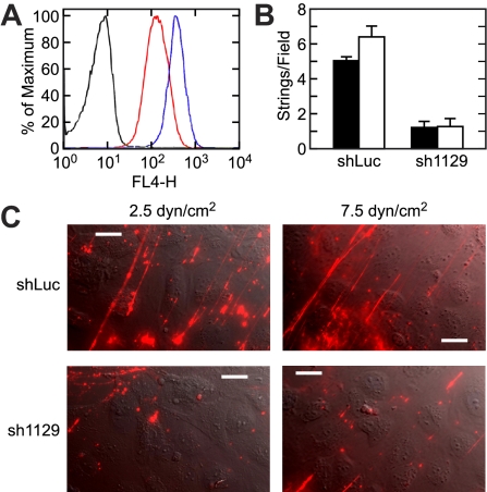
Acutely secreted von Willebrand factor (VWF) multimers adhere to endothelial cells, support platelet adhesion, and may induce microvascular thrombosis. Immunofluorescence microscopy of live human umbilical vein endothelial cells showed that VWF multimers rapidly formed strings several hundred micrometers long on the cell surface after stimulation with histamine. Unexpectedly, only a subset of VWF strings supported platelet binding, which depended on platelet glycoprotein Ib. Electron microscopy showed that VWF strings often consisted of bundles and networks of VWF multimers, and each string was tethered to the cell surface by a limited number of sites. Several approaches implicated P-selectin and integrin alpha(v)beta(3) in anchoring VWF strings. An RGDS peptide or a function-blocking antibody to integrin alpha(v)beta(3) reduced the number of VWF strings formed. In addition, integrin alpha(v) decorated the VWF strings by immunofluorescence microscopy. Furthermore, lentiviral transduction of shRNA against the alpha(v) subunit reduced the expression of cell-surface integrin alpha(v)beta(3) and impaired the ability of endothelial cells to retain VWF strings. Soluble P-selectin reduced the number of platelet-decorated VWF strings in the absence of Ca(2+) and Mg(2+) but had no effect in the presence of these cations. These results indicate that VWF strings bind specifically to integrin alpha(v)beta(3) on human endothelial cells.


Figures

Figure 1
Figure 1
Acutely secreted VWF forms extended strings under fluid shear stress. (A) Confluent HUVECs were stimulated with 100 μM histamine for 30 minutes, washed with DPBS, and fixed with 2% paraformaldehyde without permeabilization. Cell-surface VWF was stained with a polyclonal antibody (A082; Dako North America) and an Alexa Fluor 594–conjugated secondary antibody. Bar represents 10 μm. (B) HUVECs in a parallel plate flow chamber were perfused with medium 199 supplemented with 2% BSA, 100 μM histamine, and fluorescent VWF polyclonal antibody, at a shear stress of 10 dyne/cm2. Images were captured 5 minutes after the initiation of flow. Bar represents 10 μm. (C) Perfusion assays were conducted at different values of fluid shear stress. The bars indicate the numbers (mean ± SEM) of VWF strings more than 20 μm long formed 5 minutes after the initiation of flow from 10 fields per experiment. Each experiment was performed at least 3 times.
Figure 2
Figure 2
Immunoelectron microscopy of VWF strings. HUVECs were incubated with medium containing 100 μM histamine for 10 minutes with gentle rocking, washed with DPBS, and fixed with 3% paraformaldehyde. Cell-surface VWF multimers were labeled with 12-nm gold-conjugated antibody and visualized by quick-freeze deep-etch electron microscopy. Multiple VWF strands (A) form twisted bundles that sometimes bifurcate (B) and connect with one another to form networks (C). Arrows indicate branching points. Bars represent 500 nm.
Figure 3
Figure 3
A subset of VWF strings binds platelets. Perfusion assays were performed in the presence of 100 μM histamine, 108/mL fixed platelets, and fluorescent VWF polyclonal antibody. (A) A representative image of VWF strings and platelet strings for perfusion assay conducted at 2.5 dyne/cm2. Bar represents 10 μm. (B) Total fluorescent VWF strings (■) and platelet-decorated VWF strings (□) formed under a fluid shear stress of 2.5, 10, and 20 dyne/cm2 are shown as the mean plus or minus SEM from 10 fields per experiment. (C) Perfusion assays were performed at a shear stress of 2.5 dyne/cm2 in the presence or absence of 20 μg/mL antiplatelet GPIbα antibody 6D1. Total fluorescent VWF strings (■) and platelet-decorated VWF strings (□) are shown as mean plus or minus SEM from 10 fields per experiment. Each experiment was repeated at least twice.
Figure 4
Figure 4
VWF strings attach to HUVECs by discrete adhesion sites. (A) HUVECs were stimulated with histamine and exposed to laminar flow at a shear stress of 2.5 dyne/cm2. After 5 minutes, flow direction was reversed and images were captured every 30 seconds. indicate the initial flow direction (start) and the reversed flow direction (0-120 s). indicate anchorage sites for VWF strings. Bars represent 20 μm. (B) Electron micrographs of immunogold-labeled VWF strings. Samples were prepared as described in “Immunoelectron microscopy.” Arrows indicate cell extrusions in direct contact with VWF strings. Bars represent 500 nm.
Figure 5
Figure 5
P-selectin and VWF string formation. (A) Wells in an enzyme-linked immunosorbent assay plate were coated with either 20 μg/mL purified P-selectin (R&D Systems) or 1% BSA. U937 cells, which express P-selectin ligand PSGL-1, were resuspended at 106/mL, preincubated with buffer (Ctrl) or a polyclonal antibody against P-selectin (Ab) at 5 μg/mL or 10 μg/mL, and adhesion assays were performed as described in “U937 cell adhesion assay.” Results indicate the mean plus or minus SD for quadruplicate wells. (B) Perfusion assays were performed at a shear stress of 2.5 dyne/cm2. Numbers of VWF string formed under control conditions (Ctrl), with P-selectin antibody (Ab), or with purified P-selectin (P-selectin) at the indicated concentrations (μg/mL) are shown. (C) Perfusion assays were conducted in Ca2+- and Mg2+-free DPBS at a shear stress of 2.5 dyne/cm2, and the numbers of VWF strings (■) and platelet-decorated VWF strings (□) are shown as the mean plus or minus SEM from 10 fields per experiment. Cells were treated with no agonist, or with 100 μM of histamine in the absence (Ctrl) or presence of anti–P-selectin antibody (Ab) or soluble P-selectin at the indicated concentrations (μg/mL). Each experiment was performed 3 times.
Figure 6
Figure 6
Integrin αvβ3 is important for VWF string adhesion. (A) Confluent HUVECs in a flow chamber were perfused at a shear stress of 2.5 dyne/cm2 using medium 199 supplemented with 100 μM histamine, 2% BSA, and the indicated concentration of fibronectin CS-1 peptide or RGDS peptide, and the number of VWF strings formed was quantified by immunofluorescence microscopy. (B) Perfusion assays were performed similarly with the indicated concentrations of mouse IgG (mIgG) or anti-integrin αvβ3 antibody LM609, which blocks ligand binding. (C) Perfusion assays were performed similarly without (Ctrl) or with 10 μg/mL anti-integrin αvβ3 antibody LM609 or function blocking anti-integrin α5 antibody CBL497. (D) Perfusion assays were conducted without (Ctrl) or with 10 μg/mL anti-integrin αvβ3 antibody LM609 (which blocks ligand binding) or LM142 (which does not block ligand binding). (E,F) Cells were perfused with Ca2+- and Mg2+-free DPBS (E) without (No Agonist) or with 100 μM histamine in the absence (Ctrl) or presence of RGDS peptide (40 μg/mL), or (F) without (Ctrl) or with anti-integrin αvβ3 antibody LM609 (10 μg/mL), and total VWF strings (■) and platelet-decorated VWF strings (□) were quantitated. Results are shown as the mean plus or minus SEM from 10 fields per experiment. Each experiment was performed 3 times.
Figure 7
Figure 7
Integrin αv decorates acutely secreted VWF strings. Confluent HUVECs in a flow chamber were stimulated with 100 μM histamine at 2.5 dyne/cm2 shear stress for 5 minutes, fixed, and incubated with anti-VWF and anti-integrin αvβ3 antibodies as described in “Immunofluorescence microscopy.” Panels show immunofluorescence for VWF (A,D) and integrin αvβ3 (B,E) with corresponding fluorescent antibodies. Merged images are shown in panels C and F. Arrows indicate selected examples of the colocalization of VWF and integrin αvβ3. Bars represent 10 μm.
Figure 8
Figure 8
VWF string formation depends on integrin αv expression. HUVECs were infected with control shRNA lentivirus (shLuc) or integrin αv shRNA lentivirus (sh1129) and cultured for 2 weeks under selection with puromycin. (A) Fluorescence-activated cell sorter analysis was performed with control antibody on cells treated with shLuc (black trace) or with anti-integrin αvβ3 antibody LM609 on cells treated with shLuc (blue trace) or sh1129 (red trace). The level of integrin αvβ3 was reduced 70% in sh1129 cells. (B) Lentivirus-infected HUVECs were stimulated with histamine and exposed to fluid shear stress of 2.5 dyne/cm2 (■) or 7.5 dyne/cm2 (□). VWF strings were stained in situ with fluorescent anti-VWF antibody and counted. Values represent the mean plus or minus SEM from 10 fields per experiment. (C) Representative images are shown. Flow direction is from bottom left to top right. Bars represent 20 μm.

Comment in

  • Fishing for platelets.
    Cutler DF. Cutler DF. Blood. 2009 Feb 12;113(7):1397-8. doi: 10.1182/blood-2008-11-188524. Blood. 2009. PMID: 19221043

References

    1. Ruggeri ZM. The role of von Willebrand factor in thrombus formation. Thromb Res. 2007;120(Suppl 1):5–9. - PMC - PubMed
    1. Wagner DD. Cell biology of von Willebrand factor. Annu Rev Cell Biol. 1990;6:217–246. - PubMed
    1. Sadler JE. von Willebrand factor: two sides of a coin. J Thromb Haemost. 2005;3:1702–1709. - PubMed
    1. Furlan M, Robles R, Lamie B. Partial purification and characterization of a protease from human plasma cleaving von Willebrand factor to fragments produced by in vivo proteolysis. Blood. 1996;87:4223–4234. - PubMed
    1. Dong JF, Moake JL, Nolasco L, et al. ADAMTS-13 rapidly cleaves newly secreted ultralarge von Willebrand factor multimers on the endothelial surface under flowing conditions. Blood. 2002;100:4033–4039. - PubMed

Publication types

MeSH terms