

Lats2 is a negative regulator of myocyte size in the heart
- PMID: 18927464
- PMCID: PMC2775813
- DOI: 10.1161/CIRCRESAHA.108.180042
Lats2 is a negative regulator of myocyte size in the heart
Erratum in
- Circ Res. 2009 Feb 13;104(3):e27-9
Abstract
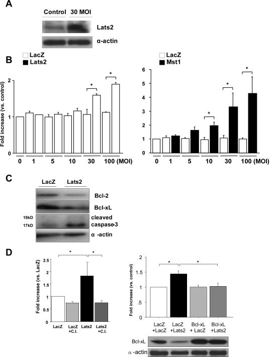
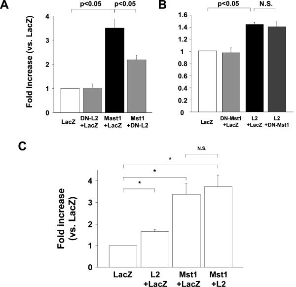
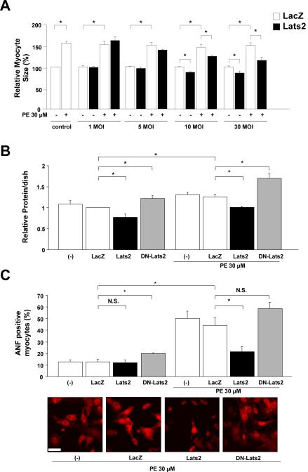
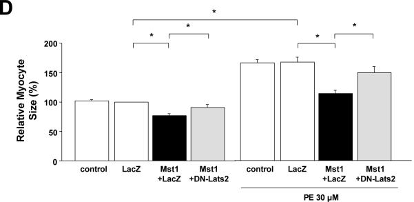
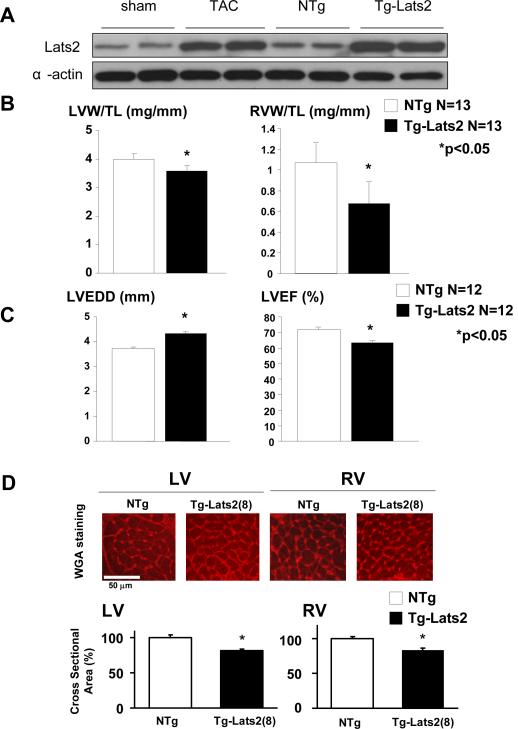
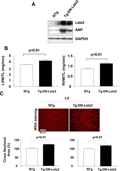
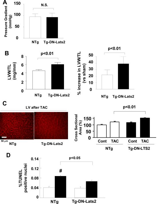
Mammalian sterile 20-like kinase (Mst)1 plays an important role in mediating apoptosis and inhibiting hypertrophy in the heart. Because Hippo, a Drosophila homolog of Mst1, forms a signaling complex with Warts, a serine/threonine kinase, which in turn stimulates cell death and inhibits cell proliferation, mammalian homologs of Warts, termed Lats1 and Lats2, may mediate the function of Mst1. We here show that Lats2, but not Lats1, dose-dependently increased apoptosis in cultured cardiac myocytes. Lats2 also dose-dependently reduced [(3)H]phenylalanine incorporation and cardiac myocyte size, whereas dominant negative Lats2 (DN-Lats2) increased them, suggesting that endogenous Lats2 negatively regulates myocyte growth. DN-Lats2 significantly attenuated induction of apoptosis and inhibition of hypertrophy by Mst1, indicating that Lats2 mediates the function of Mst1 in cardiac myocytes. Cardiac specific overexpression of Lats2 in transgenic mice significantly reduced the size of left and right ventricles, whereas that of DN-Lats2 caused hypertrophy in both ventricles. Overexpression of Lats2 reduced left ventricular systolic and diastolic function without affecting baseline levels of myocardial apoptosis. Expression of endogenous Lats2 was significantly upregulated in response to transverse aortic constriction. Overexpression of DN-Lats2 significantly enhanced cardiac hypertrophy and inhibited cardiac myocyte apoptosis induced by transverse aortic constriction. These results suggest that Lats2 is necessary and sufficient for negatively regulating ventricular mass in the heart. Although Lats2 is required for cardiac myocyte apoptosis in response to pressure overload, it was not sufficient to induce apoptosis at baseline. In conclusion, Lats2 affects both growth and death of cardiac myocytes, but it primarily regulates the size of the heart and acts as an endogenous negative regulator of cardiac hypertrophy.
Figures
References
-
- Stanger BZ. Organ size determination and the limits of regulation. Cell Cycle. 2008;7:318–324. - PubMed
-
- Beltrami AP, Barlucchi L, Torella D, Baker M, Limana F, Chimenti S, Kasahara H, Rota M, Musso E, Urbanek K, Leri A, Kajstura J, Nadal-Ginard B, Anversa P. Adult cardiac stem cells are multipotent and support myocardial regeneration. Cell. 2003;114:763–776. - PubMed
-
- Yamamoto S, Yang G, Zablocki D, Liu J, Hong C, Kim SJ, Soler S, Odashima M, Thaisz J, Yehia G, Molina CA, Yatani A, Vatner DE, Vatner SF, Sadoshima J. Activation of Mst1 causes dilated cardiomyopathy by stimulating apoptosis without compensatory ventricular myocyte hypertrophy. J Clin Invest. 2003;111:1463–1474. - PMC - PubMed
-
- Morisco C, Sadoshima J, Trimarco B, Arora R, Vatner DE, Vatner SF. Is treating cardiac hypertrophy salutary or detrimental: the two faces of Janus. Am J Physiol (Heart Circ Physiol) 2003;284:H1043–H1047. - PubMed
-
- Saucedo LJ, Edgar BA. Filling out the Hippo pathway. Nat Rev Mol Cell Biol. 2007;8:613–621. - PubMed
Publication types
MeSH terms
Substances
Grants and funding
LinkOut - more resources
Full Text Sources
Other Literature Sources
Molecular Biology Databases
Research Materials
Miscellaneous
