. 2009 Jan;27(1):138-49.
doi: 10.1634/stemcells.2008-0439.

Isolation and characterization of pluripotent human spermatogonial stem cell-derived cells

Affiliations
Free PMC article

Isolation and characterization of pluripotent human spermatogonial stem cell-derived cells

Nina Kossack et al. Stem Cells. 2009 Jan.
Free PMC article

Abstract

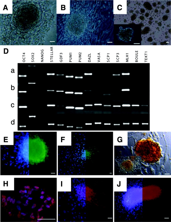
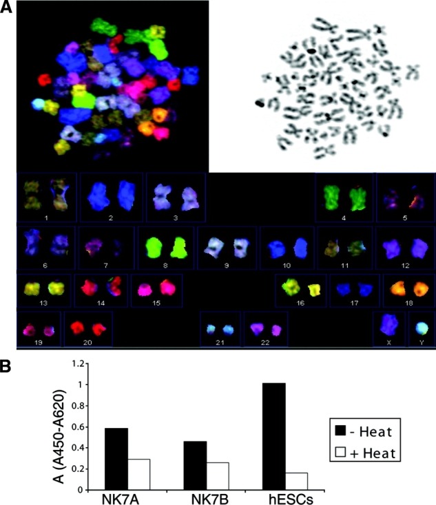
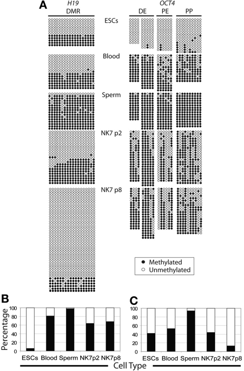
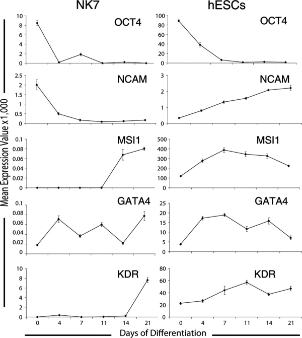
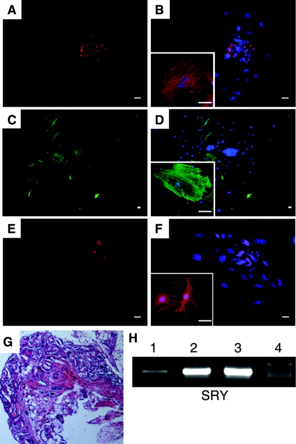
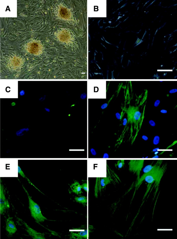
Several reports have documented the derivation of pluripotent cells (multipotent germline stem cells) from spermatogonial stem cells obtained from the adult mouse testis. These spermatogonia-derived stem cells express embryonic stem cell markers and differentiate to the three primary germ layers, as well as the germline. Data indicate that derivation may involve reprogramming of endogenous spermatogonia in culture. Here, we report the derivation of human multipotent germline stem cells (hMGSCs) from a testis biopsy. The cells express distinct markers of pluripotency, form embryoid bodies that contain derivatives of all three germ layers, maintain a normal XY karyotype, are hypomethylated at the H19 locus, and express high levels of telomerase. Teratoma assays indicate the presence of human cells 8 weeks post-transplantation but limited teratoma formation. Thus, these data suggest the potential to derive pluripotent cells from human testis biopsies but indicate a need for novel strategies to optimize hMGSC culture conditions and reprogramming.


Figures

Figure 1
Figure 1
Morphological analysis and analysis for the expression of both pluripotency- and germ cell-specific markers. (A): Light microscopy of spermatogonial stem cell colonies growing on top of a monolayer of testicular cells approximately 2 weeks after plating of the testicular cell suspension. (B): After six passages in culture. (C): Spermatogonial stem cells in suspension. (Da): Expression analysis of embryonic stem cell- and germ cell-specific markers in NK (Nina Kossack) tissue sample 7 (NK7) human multipotent germline stem cells (hMGSCs) cultured on mouse embryonic fibroblasts after two passages and seven passages. (Db, Dc): Expression analysis of NK7 cells cultured on human testicular stromal cells after 10 passages and of a commercially available testis sample. (Dd): Reverse transcriptase-polymerase chain reaction products separated by gel electrophoresis. (E–H): Immunofluorescence staining of hMGSCs at passage 8, for pluripotency markers: (E): stage-specific embryonic antigen 4. (F): TRA1–81. (G): Staining of hMGSCs at passage 8 for embryonic stem cell and germ cell-specific marker alkaline phosphatase. Shown are two colonies with different morphologies. (H): OCT4. (I): SOX2. (J): Staining of hMGSCs at passage 8 for embryonic stem cell and germ cell-specific marker DAZL. (E, F, I, J) show the colocalization of 4,6-diamidino-2-phenylindole with the corresponding pluripotency-germ cell marker on the left side and the respective marker on the right side of the image. Scale bars = 50 μm.
Figure 2
Figure 2
Human multipotent germline stem cells (hMGSCs) have a normal karyotype and express telomerase. (A): The DNA spectral karyotyping experiment of undifferentiated hMGSCs at passage 8 demonstrates a normal (46,XY) karyotype. The spectral image, the 4,6-diamidino-2-phenylindole staining, and the resulting chromosome table are shown. (B): Telomerase activity was investigated in hMGSCs at passage 6 (NK7A) and passage 8 (NK7B) using the TRAPeze ELISA. A cell extract from HSF8 hESCs served as a positive control for this experiment. The telomerase activity was calculated as average change in absorbance of the sample at 450 nm minus the absorbance at 690 nm. The black bars represent the samples without heat treatment, and the white bars represent the samples with heat treatment. Abbreviations: A, absorbance; hESC, human embryonic stem cell.
Figure 3
Figure 3
Bisulfite sequencing of the OCT4 promoter region and the DMR, located upstream of the H19 promoter. (A): Methylation profile of the human OCT4 and H19 genes. Each row of circles represents a single cloned allele, and each circle represents a single CpG site (white circles, nonmethylated cytosine; black circles, methylated cytosine). Bisulfite-modified DNA from H9 human embryonic stem cells, human multipotent germline stem cells (NK7) at p2 and p8, blood cells, and sperm cells were analyzed. (B): Percentages of methylated and unmethylated clones present in the DMR of the H19 gene, as portrayed in (A) above. White bars indicate the percentages of unmethylated clones, and black bars indicate the percentages of methylated clones for each cell type as shown along the x-axis. (C): Percentages of methylated and unmethylated clones present in the promoter region of the OCT4 gene, as portrayed in (A). White bars indicate the percentages of unmethylated clones, and black bars indicate the percentages of methylated clones for each cell type as shown along the x-axis. Abbreviations: DE, distal enhancer; DMR, differentially methylated region; NK7, NK (Nina Kossack) tissue sample 7; p, passage; PE, proximal enhancer; PP, proximal promoter.
Figure 4
Figure 4
In vitro differentiation of human multipotent germline stem cells (hMGSCs). hMGSCs and H9 human embryonic stem cells were spontaneously differentiated in vitro over a period of 21 days. RNA was isolated at six time points, and TaqMan reverse transcriptase-polymerase chain reaction was performed to quantify the expression of the pluripotency marker OCT4 and the somatic genes NCAM, MLH1 (ectoderm marker), GATA4 (endoderm marker), and KDR (mesoderm marker). Expression values were calculated as previously described [35] and were normalized using GAPDH as a reference. Abbreviation: NCAM, neural cell adhesion molecule.
Figure 5
Figure 5
Immunofluorescence staining of day 7 differentiated human multipotent germline stem cells and teratoma analysis. Attached EBs were assessed for protein expression as shown in (A-F). (A): von Willebrand factor (VWF). (B): VWF with 4,6-diamidino-2-phenylindole (DAPI) overlay. (C): α-Smooth muscle actin (ASMA). (D): ASMA with DAPI overlay. (E): Nestin (NES). (F): NES with DAPI overlay. Scale bars = 50 μm. (G): Analysis of teratomas 8 weeks post-transplantation. Shown is a representative section of the NK (Nina Kossack) tissue sample 7 (NK7) cell graft after 8 weeks of in vivo differentiation. (H): Expression analysis of the human SRY gene was performed using genomic DNA isolated from an NK7 paraffin-embedded tissue slide (lane 1), NK7 genomic DNA (lane 2), sperm genomic DNA (lane 3), and female mouse genomic DNA as template (lane 4). Shown are polymerase chain reaction products separated by gel electrophoreses. The fragment amplified with the SRY primers had a size of 350 base pairs. Abbreviation: SRY, sex determining region Y.
Figure 6
Figure 6
Immunofluorescence staining after 6 weeks of induced neural differentiation. Human multipotent germline stem cells were cultured on mouse embryonic fibroblasts in human embryonic stem cell media (A) and were plated onto gelatin (B) prior to the differentiation experiment. Cells were stained for nestin (C) before the treatment and following induced differentiation (D). Differentiated cells were also stained for the ectodermal markers microtubule-associated protein 2 (E) and β-tubulin III (F). Scale bars = 50 μm.

References

    1. Martin G. Isolation of a pluripotent cell line from early mouse embryos cultured in medium conditioned by teratocarcinoma stem cells. Proc Natl Acad Sci U S A. 1981;78:7634–7638. - PMC - PubMed
    1. Kluin PM, de Rooij DG. A comparison between the morphology and cell kinetics of gonocytes and adult type undifferentiated spermatogonia in the mouse. Int J Androl. 1981;4:475–493. - PubMed
    1. Rossant J. Stem cells from the mammalian blastocyst. Stem Cells. 2001;19:477–482. - PubMed
    1. Evans M, Hunter S. Source and nature of embryonic stem cells. C R Biol. 2002;325:1003–1007. - PubMed
    1. Buehr M, Smith A. Genesis of embryonic stem cells. Philos Trans R Soc Lond B Biol Sci. 2003;358:1397–1402. - PMC - PubMed

Publication types

MeSH terms