. 2008;3(11):1736-50.
doi: 10.1038/nprot.2008.174.

DNA cleavage assay for the identification of topoisomerase I inhibitors

Affiliations

DNA cleavage assay for the identification of topoisomerase I inhibitors

Thomas S Dexheimer et al. Nat Protoc. 2008.

Abstract

The inhibition of DNA topoisomerase I (Top1) has proven to be a successful approach in the design of anticancer agents. However, despite the clinical successes of the camptothecin derivatives, a significant need for less toxic and more chemically stable Top1 inhibitors still persists. Here, we describe one of the most frequently used protocols to identify novel Top1 inhibitors. These methods use uniquely 3'-radiolabeled DNA substrates and denaturing polyacrylamide gel electrophoresis to provide evidence for the Top1-mediated DNA cleaving activity of potential Top1 inhibitors. These assays allow comparison of the effectiveness of different drugs in stabilizing the Top1-DNA intermediate or cleavage (cleavable) complex. A variation on these assays is also presented, which provides a suitable system for determining whether the inhibitor blocks the forward cleavage or religation reactions by measuring the reversibility of the drug-induced Top1-DNA cleavage complexes. This entire protocol can be completed in approximately 2 d.


Figures

Figure 1 |
Figure 1 |
Flow diagram to illustrate the major steps of the protocol.
Figure 2 |
Figure 2 |
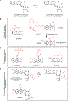
Structure and mechanism of the Top1 cleavage complex (Top 1Cc) trapping by a specific Top1 inhibitor (indenoisoquinoline derivative),,. (a,b) Top1 nicking–closing reaction,. (a) Top1 is generally bound noncovalently to DNA. The Top1 catalytic tyrosine (Y723 for human nuclear Top1) is represented in red (Y). (a to b) Top1 cleaves one strand of the duplex, as it forms a covalent phosphodiester bond between the catalytic tyrosine and the 3′-DNA terminus. The other DNA terminus is a 5′-hydroxyl (OH). (b) The Top 1Cc allows rotation of the 5′-terminus around the intact strand, which relaxes DNA supercoiling (purple dotted circle with arrowhead). (b to a) Following DNA relaxation, Top1 religates the DNA. Under normal conditions, the religation (closing) reaction rate constant is much higher than the cleavage (nicking) rate constant. More than 90% of the Top1–DNA complexes are noncovalent. (c) Top1 inhibitors (green and red polygon; structures shown in Figs. 3 and 4) trap Top 1Cc by the binding of one drug molecule at the enzyme–DNA interface between the base pairs flanking the Top1-mediated DNA cleavage site (by convention positions −1 and +1). (d,e) Lateral views of a Top 1Cc trapped by an indenoisoquinoline derivative (drug shown in green and red; DNA in dark blue; Top1 in yellow),,. (d) Top1 is shown in a surface view to represent the depth of the indenoisoquinoline binding pocket. (e) Top1 is represented in ribbon diagram to allow visualization of the catalytic tyrosine (Y; red) and to show the drug intercalation between the −1 and +1 base pairs,,.
Figure 3 |
Figure 3 |
Structures of camptothecin derivatives. (a) The active camptothecin alkaloid is the 20-(S) enantiomer (as shown with the 20-hydroxyl above the plane). Camptothecin is spontaneously and reversibly converted to its carboxylate derivative, which binds serum albumin and is inactive against Top1. (b) The two camptothecins approved for cancer chemotherapy are topotecan and irinotecan. Both are water soluble, but irinotecan is a prodrug and needs to be converted to SN-38. (c) Camptothecin derivatives in clinical trials. Gimatecan can be given orally. (d) Two strategies have been used to overcome the inactivation of camptothecins by E-ring opening. An additional methylene group yields homocamptothecins with a more stable E-ring,. However, once converted to carboxylates, homocamptothecins cannot reverse to the active E-ring-closed form. Removal of the oxygen atom in the E-ring yields keto derivatives that cannot undergo E-ring opening. S39625 has just begun clinical trials. Substitutions on the camptothecin structure are shown in red.
Figure 4 |
Figure 4 |
Structures of the three chemical families of noncamptothecin Top1 inhibitors. The indolocarbazole derivative, edotecarin, is in Phase II clinical trials. Two indenoisoquinolines are in preclinical development,. ARC-111 (topovale) (or one of its derivatives) is also in preclinical development,.
Figure 5 |
Figure 5 |
Cartoon illustrating the advantages of using a 3′-end-labeled DNA substrate. (a) Example of theoretical products that are produced following the Top1-mediated DNA cleavage reaction in the presence of a Top1 inhibitor. Blue circles correspond to the 3′-end label. (b) Migration of the denatured DNA fragments on a denaturing polyacrylamide gel. Labeled DNA fragments, which are visible after the PhosphorImager scan, are shown in black. Unlabeled DNA fragments are shown in gray.
Figure 6 |
Figure 6 |
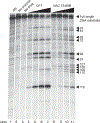
Top1-mediated DNA cleavage assay. Equal amounts of recombinant Top1 were incubated with the 3′-end-labeled DNA substrate in the presence (CPT and NSC 724998) or absence of drug. The reaction products were resolved on a denaturing polyacrylamide gel. Lane 1, Maxam–Gilbert AG sequencing reaction; lane 2, negative enzyme control; lane 3, negative inhibitor control; lanes 4–7, 0.01, 0.1, 1 and 10 μM of CPT; lanes 8–11, 0.01, 0.1, 1 and 10 mM of NSC 724998. Arrows and numbers indicate the sites of DNA cleavage.
Figure 7 |
Figure 7 |
Top1–DNA cleavage complex reversal assay. After 20 min of incubation with 0.1 or 10 μM of CPT or NSC 724998, respectively, 0.35 M NaCl was added to induce the reversal of the Top1 cleavage complexes. Aliquots were taken and reactions were terminated at various time points: 0, immediately before NaCl addition (lanes 4 and 11), 0.5 min (lanes 5 and 12), 1 min (lanes 6 and 13), 2 min (lanes 7 and 14), 5 min (lanes 8 and 15), 10 min (lanes 9 and 16) and 20 min (lanes 10 and 17).

References

    1. Kohlhagen G, Paull K, Cushman M, Nagafufuji P & Pommier Y Protein-linked DNA strand breaks induced by NSC 314622, a non-camptothecin topoisomerase I poison. Mol. Pharmacol. 54, 50–58 (1998). - PubMed
    1. Strumberg D et al. Synthesis of cytotoxic indenoisoquinoline topoisomerase I poisons. J. Med. Chem. 42, 446–457 (1999). - PubMed
    1. Antony S et al. Cellular Topoisomerase I Inhibition and antiproliferative activity by MJ-III-65 (NSC 706744), an indenoisoquinoline topoisomerase I poison. Mol. Pharmacol. 67, 523–530 (2005). - PubMed
    1. Ioanoviciu A et al. Synthesis and mechanism of action studies of a series of norindenoisoquinoline topoisomerase I poisons reveal an inhibitor with a flipped orientation in the ternary DNA-enzyme-inhibitor complex as determined by X-ray crystallographic analysis. J. Med. Chem. 48, 4803–4814 (2005). - PubMed
    1. Staker BL et al. Structures of three classes of anticancer agents bound to the human topoisomerase I-DNA covalent complex. J. Med. Chem. 48, 2336–2345 (2005). - PubMed

Publication types

MeSH terms

Substances