. 2008;3(10):e3441.
doi: 10.1371/journal.pone.0003441. Epub 2008 Oct 17.

DeltaNp63 is essential for epidermal commitment of embryonic stem cells

Affiliations

DeltaNp63 is essential for epidermal commitment of embryonic stem cells

Alain Medawar et al. PLoS One. 2008.

Abstract

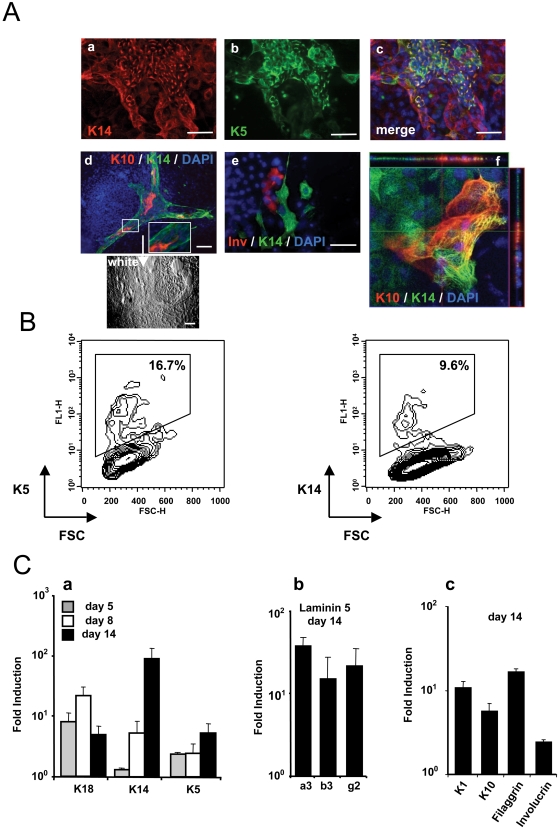
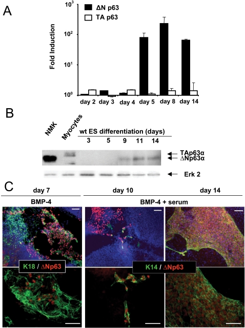
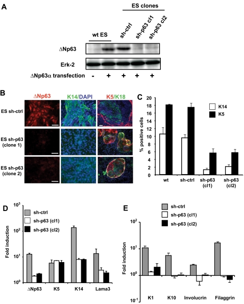
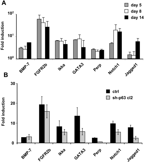
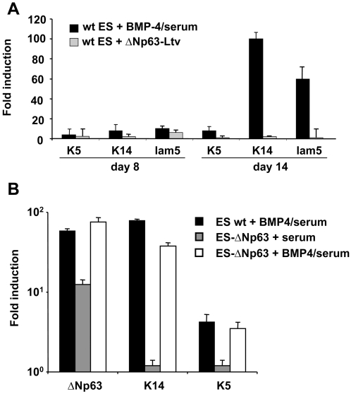
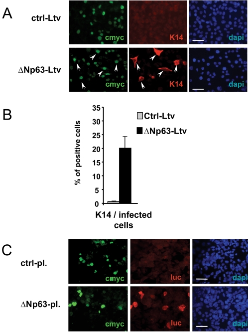
In vivo studies have demonstrated that p63 plays complex and pivotal roles in pluristratified squamous epithelial development, but its precise function and the nature of the isoform involved remain controversial. Here, we investigate the role of p63 in epithelial differentiation, using an in vitro ES cell model that mimics the early embryonic steps of epidermal development. We show that the DeltaNp63 isoform is activated soon after treatment with BMP-4, a morphogen required to commit differentiating ES cells from a neuroectodermal to an ectodermal cell fate. DeltaNp63 gene expression remains high during epithelial development. P63 loss of function drastically prevents ectodermal cells to commit to the K5/K14-positive stratified epithelial pathway while gain of function experiments show that DeltaNp63 allows this commitment. Interestingly, other epithelial cell fates are not affected, allowing the production of K5/K18-positive epithelial cells. Therefore, our results demonstrate that DeltaNp63 may be dispensable for some epithelial differentiation, but is necessary for the commitment of ES cells into K5/K14-positive squamous stratified epithelial cells.


Conflict of interest statement

Competing Interests: The authors have declared that no competing interests exist.

Figures

Figure 1
Figure 1. Epidermal differentiation of murine ES cells.
(A) Appearance of K5+/K14+ and involucrin-positive epidermal cells from mouse ES cells treated with BMP-4 and serum. The differentiated cells were immunostained for cytokeratin K14, K5, K10 or involucrin at day 14 (panels a–e). Scale bars = 50 µm. Confocal analysis of differentiated cells (at day 14) (panel f). (B) Flow cytofluorimetry analysis of K5 and K14 on differentiated cells at day 14 of culture. The graphs represent the fluorescence intensity for the indicated proteins plotted against the cell size (forward scatter, FSC). The percentages of positive cells among the gated populations are indicated on graphs. (C) Gene expression was analyzed by real-time RT-PCR, for K18, K5, and K14 at the indicated differentiation time points (panel a), for laminin-5 chains at day 14 (panel b), and for K1, K10, filaggrin and involucrin at day 14 (panel c). The value for each gene was normalized to untreated control ES cultures, and represents the average of three independent experiments±sd.
Figure 2
Figure 2. Expression of ΔNp63 during epidermal differentiation of murine ES cells.
(A) Real-time RT-PCR analysis for ΔN- and TA-p63 isoform gene expression at the indicated differentiation time points. The value for each gene was normalized to untreated control ES cultures, and represents the average of three independent experiments±sd. (B) Immunoblots of endogenous p63 isoforms in normal mouse keratinocytes (NMK), myocytes and ES cells during 3, 5, 9, 11 and 14 days of differentiation, as revealed by anti-p63 and anti-Erk2 (as loading control) antibodies. Normal mouse keratinocytes (known to express endogenous ΔNp63 isoforms) and differentiating myocytes (which have been shown to express the TAp63 isoform, [24]) served as molecular weight references. (C) Immunofluorescence detection of ΔNp63 during ES cell differentiation. The differentiating cells were immunostained for K18 or K14 (green) and for ΔNp63 (red). Dapi staining is shown in blue. Scale bars = 50 µm.
Figure 3
Figure 3. p63 is required for epithelial differentiation of ES cells.
Stable ES cell clones in which ΔNp63 gene expression has been inhibited with shRNA, were produced and induced to differentiate into the epidermal fate. (A) Analysis of ES clones transfected with a control pSuper sh-RNA (ctrl) or a pSuper sh-RNA specific for p63 (clones sh-1, sh-2). Wild type ES cells and stable clones were transiently transfected with a ΔNp63-expressing vector and analyzed 48 h later by Western blot with anti-ΔNp63 and anti-Erk2 (as loading control) antibodies. The two p63 sh-RNA clones displayed a strong inhibition of the ectopic ΔNp63 expression while the pSuper control was unable to regulate it. (B) Immunofluorescence staining for the indicated proteins in control and sh-p63 ES cell clones at day 14 of differentiation. Dapi staining is shown in blue. Bars = 50 µm. (C) Percentages of K14- or K5-positive cells differentiating from wt-ES cells, control-sh or p63-specific sh ES clones at day 14 determined by flow cytofluorometric analysis. (D) Real-time RT-PCR analysis at day 14 of differentiation for ΔNp63, K5, K14 and Lama3 (left panel) and for K1, K10, filaggrin and involucrin (right panel) genes. ES cell clones expressing either a control- or p63-specific shRNA were tested.
Figure 4
Figure 4. Time course of relative mRNA expression of several p63-regulated genes.
(A) Real-time RT-PCR analysis for p63-targets genes at 3 differentiation time points, normalized to untreated control ES cultures. The data represents the average of three independent experiments±sd. (B) Relative mRNA expression for different p63-regulated genes. (A) Real-time RT-PCR analysis for p63-target genes at 3 differentiation time points, normalized to undifferentiated ES cells at day 0. (B) Real-time RT-PCR analysis for p63-target genes in control and sh-p63 cl2 ES cells at day 14 normalized to undifferentiated WT ES cells at day. The data represents the average of three experiments±sd.
Figure 5
Figure 5. Ectopic expression of ΔNp63 does not induce efficient stratified epithelial commitment of ES cells.
(A) Differentiating ES cells were either treated with BMP-4 (at day 3) or transduced (at day 4) with lentiviruses expressing either ΔNp63 or irrelevant proteins (red tomato or Pax6) as negative controls. Real-time RT-PCR analysis for K5, K14 and laminin-5 gene expression was performed at days 5, 8 and 14 of ES cell differentiation. The value for each gene was normalized to either untreated control cultures for BMP-4-treated ES cells or red tomato transduced control cultures for p63-transduced ES cells. The values represent an average of three determinations±sd. (B) Wild type ES or ES cells stably expressing ΔNp63 were induced to differentiate with and without BMP-4. Real-time RT-PCR analysis for K5, K14 and p63 gene expression was performed at day 14. The value for each gene was normalized to untreated wild type ES cells at day 14. The values represent an average of three determinations±sd.
Figure 6
Figure 6. ES-derived ectodermal cells differentiate into K14/K5-positive epidermal cells upon ΔNp63 ectopic expression.
An ectodermal K8+/K18+ cell line was derived by serial clonal dilutions of differentiating ES cells after BMP-4 treatment. (A) Immunofluorescence staining of the ES-derived ectodermal cell line 48 h after infection with a lentivirus expressing c-myc-tagged-ΔNp63. Control cells were transduced with a control c-myc-tagged-pax6 lentivirus. Nuclei are stained with DAPI. Bars = 50 µm. (B) Percentages of K14-positive cells amongst transduced cells 48 h after infection, as determined by immunofluorescence staining of c-myc and K14-positive cells. (C) Immunofluorescence staining of the ES-derived ectodermal cell line 48 h after cotransfection of c-myc-tagged-ΔNp63 and a p63-responsive reporter luciferase construct. Positive cells were detected with c-myc and luciferase antibodies. Nuclei are stained with DAPI. Bars = 50 µm.

References

    1. Yang A, Kaghad M, Wang Y, Gillett E, Fleming MD, et al. p63, a p53 homolog at 3q27-29, encodes multiple products with transactivating, death-inducing, and dominant-negative activities. Mol Cell. 1998;2:305–316. - PubMed
    1. Mills AA, Zheng B, Wang XJ, Vogel H, Roop DR, et al. p63 is a p53 homologue required for limb and epidermal morphogenesis. Nature. 1999;398:708–713. - PubMed
    1. Yang A, Schweitzer R, Sun D, Kaghad M, Walker N, et al. p63 is essential for regenerative proliferation in limb, craniofacial and epithelial development. Nature. 1999;398:714–718. - PubMed
    1. Yang A, Kaghad M, Caput D, McKeon F. On the shoulders of giants: p63, p73 and the rise of p53. Trends Genet. 2002;18:90–95. - PubMed
    1. Pellegrini G, Dellambra E, Golisano O, Martinelli E, Fantozzi I, et al. p63 identifies keratinocyte stem cells. Proc Natl Acad Sci U S A. 2001;98:3156–3161. - PMC - PubMed

Publication types