

Evidence that molecular changes in cells occur before morphological alterations during the progression of breast ductal carcinoma
- PMID: 18928525
- PMCID: PMC2614523
- DOI: 10.1186/bcr2157
Evidence that molecular changes in cells occur before morphological alterations during the progression of breast ductal carcinoma
Abstract
Introduction: Ductal carcinoma in situ (DCIS) of the breast includes a heterogeneous group of preinvasive tumors with uncertain evolution. Definition of the molecular factors necessary for progression to invasive disease is crucial to determining which lesions are likely to become invasive. To obtain insight into the molecular basis of DCIS, we compared the gene expression pattern of cells from the following samples: non-neoplastic, pure DCIS, in situ component of lesions with co-existing invasive ductal carcinoma, and invasive ductal carcinoma.
Methods: Forty-one samples were evaluated: four non-neoplastic, five pure DCIS, 22 in situ component of lesions with co-existing invasive ductal carcinoma, and 10 invasive ductal carcinoma. Pure cell populations were isolated using laser microdissection. Total RNA was purified, DNase treated, and amplified using the T7-based method. Microarray analysis was conducted using a customized cDNA platform. The concept of molecular divergence was applied to classify the sample groups using analysis of variance followed by Tukey's test.
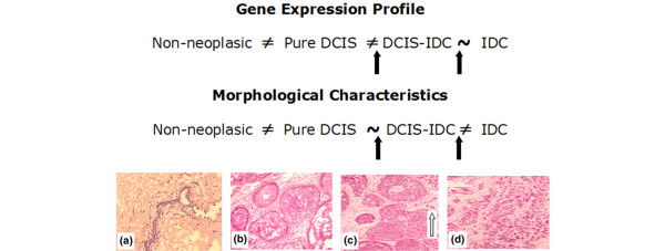
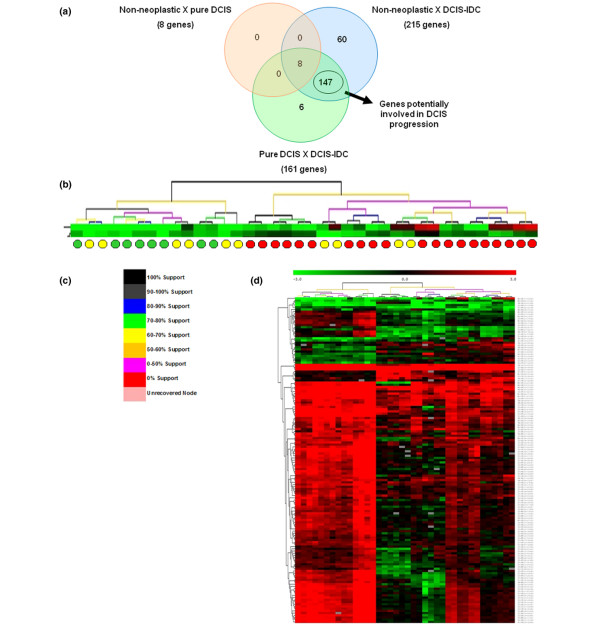
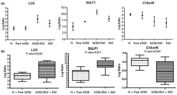
Results: Among the tumor sample groups, cells from pure DCIS exhibited the most divergent molecular profile, consequently identifying cells from in situ component of lesions with co-existing invasive ductal carcinoma as very similar to cells from invasive lesions. Additionally, we identified 147 genes that were differentially expressed between pure DCIS and in situ component of lesions with co-existing invasive ductal carcinoma, which can discriminate samples representative of in situ component of lesions with co-existing invasive ductal carcinoma from 60% of pure DCIS samples. A gene subset was evaluated using quantitative RT-PCR, which confirmed differential expression for 62.5% and 60.0% of them using initial and partial independent sample groups, respectively. Among these genes, LOX and SULF-1 exhibited features that identify them as potential participants in the malignant process of DCIS.
Conclusions: We identified new genes that are potentially involved in the malignant transformation of DCIS, and our findings strongly suggest that cells from the in situ component of lesions with co-existing invasive ductal carcinoma exhibit molecular alterations that enable them to invade the surrounding tissue before morphological changes in the lesion become apparent.
Figures

References
-
- Porter D, Lahti-Domenici J, Keshaviah A, Bae YK, Argani P, Marks J, Richardson A, Cooper A, Strausberg R, Riggins GJ, Schnitt S, Gabrielson E, Gelman R, Polyak K. Molecular markers in ductal carcinoma in situ of the breast. Mol Cancer Res. 2003;1:362–375. - PubMed
-
- Zhuang Z, Merino MJ, Chuaqui R, Liotta LA, Emmert-Buck MR. Identical allelic loss on chromosome 11q13 in microdissected in situ and invasive human breast cancer. Cancer Res. 1995;55:467–471. - PubMed
Publication types
MeSH terms
Substances
LinkOut - more resources
Full Text Sources
Other Literature Sources
Medical
Molecular Biology Databases
