. 2008 Dec 10;314(20):3724-36.
doi: 10.1016/j.yexcr.2008.09.019. Epub 2008 Oct 7.

The complexity of signaling in host-pathogen interactions revealed by the Toxoplasma gondii-dependent modulation of JNK phosphorylation

Affiliations

The complexity of signaling in host-pathogen interactions revealed by the Toxoplasma gondii-dependent modulation of JNK phosphorylation

John C Carmen et al. Exp Cell Res. .

Abstract

The inhibition of apoptosis by Toxoplasma gondii is governed by its modulation of several signaling cascades including the NFkappaappaB and JNK pathways. This is evident in the dysregulation of JNK activation following treatment with UV and TNFalpha, both apoptogenic stimuli. Infection-mediated interference with the JNK cascade was found to be highly reproducible in HeLa cells. In light of emerging evidence regarding cross talk between the JNK and NFkappaB cascades, we examined the impact of infection in wild type and RelA/p65-/- mouse embryonic fibroblasts (MEF). Remarkably, parasite infection failed to significantly impact both UV and TNFalpha-mediated JNK phosphorylation in both cell lines suggesting a cell type specific effect. Furthermore siRNA-mediated knockdown of RelA/p65 failed to impact the parasite mediated effects on stimulus dependent activation of JNK in HeLa cells. Finally, the infection mediated suppression of JNK phosphorylation in HeLa cells did not result in decreased JNK kinase activity. Rather, the reduced levels of phospho-JNK in infected cells correlated with increased phosphatase activity noted by the partial rescue of the phenotype following treatment with okadaic acid. Taken together the results indicate that manipulation of the JNK pathway does not involve NFkappaB and is furthermore not a central component of the parasite enforced block of apoptosis. It further highlights the complexity of these systems and the danger of extrapolating results both within and across pathogen-host cell systems based on limited studies.


Figures
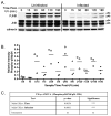
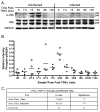
Fig. 1
Fig. 1. JNK activation is inhibited in T. gondii-infected HeLa cells following exposure to UV
(A) A representative immunoblot of uninfected and T. gondii infected HeLa cells following exposure to UV irradiation and harvested at the time points indicated. JNK-phosphorylation (PJNK) occurs within 15 minutes post-UV and is sustained for the duration of the experiment in uninfected HeLa cells. This response is markedly inhibited both with regard to the extent of JNK phosphorylation and its kinetics in cells infected with T. gondii. Total JNK (JNK) and calnexin serve as loading controls. (B) Densitometric analysis representing the level of phospho-p54 JNK relative to total JNK in three independent experiments depicts the range of values inherent in such experiments. Relative phospho-p54 levels in uninfected cells (circle) versus infected cells (diamond) confirm a marked inhibition of mean phospho-p54 JNK accumulation (horizontal line) in parasite infected cells relative to uninfected cells. Densitometric analysis was performed as described in the materials and methods. (C) Statistical analysis using two way ANOVA indicates highly significant effects for both time post UV and infection. Time and infection as variables however do not exhibit significant interaction. Significance: p values <0.01 are considered highly significant (***), while p values between 0.01 and 0.05 are considered significant (**) and p-values > 0.05 are deemed not significant (ns).
Fig. 2
Fig. 2. JNK activation is inhibited in T. gondii-infected HeLa cells following exposure to TNFα
(A) A representative immunoblot of uninfected and T. gondii infected HeLa cells following exposure to TNFα and harvested at the time points indicated. JNK-phosphorylation (PJNK) occurs transiently, peaking at 30 minutes post exposure. This response is markedly inhibited in cells infected with T. gondii although the kinetics of the response do not appear to be impacted. Total JNK (JNK) and calnexin serve as loading controls. (B) Densitometric analysis representing the level of phospho-p54 JNK relative to total p54 JNK in four independent experiments depicts the range of values inherent in such experiments. Relative phospho-p54 levels in uninfected cells (open circle) versus infected cells (diamond) confirm a marked inhibition of mean phospho-p54 JNK accumulation (horizontal line) in parasite infected cells relative to uninfected cells. Densitometric analysis was performed as described in the materials and methods. (C) Statistical analysis using 2 way ANOVA indicate highly significant effects for both time post-TNFα exposure and infection. Time and infection do not exhibit significant interaction. Significance: p values of 0.01 and under are considered highly significant (***), p values between 0.01 and 0.05 are considered significant (**) while those above 0.05 are not significant (ns).
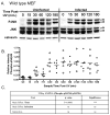
Fig. 3
Fig. 3. Phospho-JNK levels are not affected by T. gondii infection in wild type mouse embryonic fibroblasts and p65−/− cells following UV exposure
(A) A representative immunoblot of uninfected and T. gondii-infected wild type mouse embryonic fibroblasts following exposure to UV irradiation and harvested at the time points indicated. JNK-phosphorylation (PJNK) occurs within 15 minutes post UV and is sustained for the duration of the experiment in uninfected MEF cells similar to the pattern observed in HeLa cells. This response is fundamentally identical with regard to the extent of JNK phosphorylation and its kinetics in cells infected with T. gondii. Total JNK (JNK) and calnexin serve as loading controls. (B) Densitometric of phospho-p54 JNK levels relative to total JNK in six independent experiments depicts the range of values inherent in such experiments. Relative phospho-p54 levels in uninfected cells (circle) versus infected cells (diamond) confirm an absence of inhibition of mean phospho-p54JNK accumulation (horizontal line) in parasite-infected cells relative (diamond) to uninfected cells (circle). Densitometric analysis was performed as described in the materials and methods (C) Statistical analysis using two-way ANOVA indicate a highly significant effect for time post-UV but not for infection. Time and infection do not exhibit significant interaction. The identical experiment to that performed with wild type MEF was replicated in p65−/− fibroblasts. Results indicate the absence of any significant inhibition in relative phospho-p54 JNK levels. (E) Densitometric analysis of six independent experiments confirms the broad range of responses and the lack of apparent differences in the mean levels of phospho-p54 JNK comparing uninfected (circle) and infected (diamond) p65−/− cells. (F) Two way ANOVA analysis reveals a highly significant effect for time post-UV but not infection. Significance: p values of 0.01 and lower are considered highly significant (***), p values between 0.01 and 0.05 are considered significant (**) while those of above 0.05 are not significant (ns).
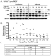
Fig. 4
Fig. 4. Phospho-JNK levels are not affected by T. gondii infection in wild type mouse embryonic fibroblasts and p65−/− cells following TNFα exposure
(A) A representative immunoblot of uninfected and T. gondii infected wild type mouse embryonic fibroblasts following exposure to TNFα and harvested at the time points indicated. JNK-phosphorylation (PJNK) occurs within 15 minutes post-UV and is transient with a pattern similar to that seen in HeLa cells. This response is fundamentally identical with regard to the extent of JNK phosphorylation and its kinetics in cells infected with T. gondii. Total JNK (JNK) and calnexin serve as loading controls. (B) Densitometric analysis representing the level of phospho-p54 JNK relative to total JNK in four independent experiments depicts the range of values inherent in such experiments. Relative phospho-p54 levels in uninfected cells (circle) versus infected cells (diamond) confirm an absence of inhibition of mean phospho-p54 JNK accumulation (horizontal line) in parasite infected cells relative (diamond) to uninfected cells (circle). Densitometric analysis was performed as described in the Materials and methods (C) Statistical analysis using two way ANOVA indicate a modest effect for time post-treatment but not for infection. Time and infection do not exhibit significant interaction.. The identical experiment to that performed with wild type MEF was replicated in p65−/− fibroblasts. Results indicate the absence of any significant inhibition in relative phospho-p54JNK levels. (E) Densitometric analysis of six independent experiments confirms the broad range of responses and the lack of apparent differences in the mean levels of phospho-p54 JNK comparing uninfected (circle) and infected (diamond) p65−/− cells. (F) Two-way ANOVA analysis reveals a highly significant effect for time post-UV but not infection. Significance: p values of 0.01 and lower are considered highly significant (***), p values between 0.01 and 0.05 are considered significant (**) while those above 0.05 are not significant (ns).
Fig. 5
Fig. 5. Depletion of RelA/p65 in HeLa cells does not affect the parasite mediated inhibition of JNK-phosphorylation in response to UV
(A) Immunofluorescence analysis of untransfected HeLa cells reveals high levels of p65 in the cytoplasm relative to cells transfected with p65-targeting siRNA. Images were acquired under identical exposure conditions. Nuclei in the cells are labeled with Hoescht dye (blue). (B) Depletion of p65 recorded by immunoblot analysis comparing mock transfected cells (siGLO reagent) to target siRNA (p65) transfected cells. Aliquots of cells used in the experiments to examine the effect on JNK phosphorylation post-UV account for the different time points in the immunoblot. Note that UV exposure (comparison of t=0 relative to other time points) results in the apparent degradation of p65 selectively in infected siRNA transfected cells. (C) Immunoblot analysis confirms the effect of UV on JNK-phosphorylation as well as the inhibition of this effect in infected mock-transfected cells. ANOVA analysis indicated highly significant effects for time (p=0.0057) and infection (p=0.0138) with no interaction between these factors for mock treated cells. (D) siRNA mediated depletion of p65 (p65KD) does not have any effect on either the kinetics or temporal profile of JNK-phosphorylation in uninfected HeLa cells. ANOVA analysis confirms highly significant effects for time (p=0.0054) and infection (p=0.0013) with no interaction between these factors following p65 depletion. In addition, the capacity of T. gondii to affect JNK phosphorylation in response to UV is not affected by the depletion of p65. (E,F) Side by side comparison of mock transfected (M) vs p65 knock down (p65KD) HeLa cells reveals no significant effect on JNK phosphorylation in either uninfected (E) or infected (F) cells. Error bars represent the standard error of the mean from four independent experiments at each time point examined.
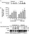
Fig. 6
Fig. 6. T. gondii-dependent inhibition of JNK phosphorylation in HeLa cells does not correlate with a decrease in the level of JNK activity
(A) The phosphorylation of cJUN by immunoprecipitated JNK following activation with UV was used as an indicator of JNK activity. Following the incubation of the immunoprecipitate with 32P-labeled ATP and recombinant cJUN, the phosphorylated cJUN was resolved via SDS-PAGE and visualized using a phosphorimager. A representative autoradiogram indicates UV-dependent activation JNK activity in both uninfected and infected cells. (B) Densitometric analysis was performed of three independent experiments. The analysis of 32P-cJUN levels confirmed that infection failed to decrease JNK activity but rather prolonged it. ANOVA analysis revealed no significant effect for time post UV or infection (data not shown). Error bars represent standard error of the mean from three independent experiments. (C) The absence of inhibition of JNK kinase activity as measured by cJUN phosphorylation following the immunoprecipitation of JNK does not correlate with the inhibition of phospho-JNK levels seen by immunoblot in an aliquot of the sample used for the immunoprecipitation.
Fig. 7
Fig. 7. Effect of inhibition of phosphatase activity using okadaic acid on phospho-JNK levels
HeLa cells were pre-treated with okadaic acid (100 nM) or the carrier control for 2 hr prior to UV irradiation (100 mJ cm−2). Cells were returned to either prewarmed treatment media or carrier control containing media immediately following irradiation and the kinetics of PJNK accumulation determined. (A,B) Treatment of uninfected cells with okadaic acid failed to increase the levels of phospho-JNK relative to the carrier control. Infected cells treated with both carrier control and okadaic acid exhibit lower levels of phospho-JNK compared to treatment and time matched uninfected cells. However, comparison of phospho-JNK levels between control and okadaic acid treated cells suggest a potential increase in relative phospho-JNK levels. (C,D) Densitometric analysis compiling the results from three independent experiments confirm the absence of any effect from okadaic acid in uninfected cells except possible a marginally increased loss of signal in okadiac acid treated cells. In contrast, treatment of infected cells with okadaic acid results in a significant increase in the levels of phospho-JNK relative to total JNK which are comparable to those seen in uninfected cells. This suggests an okadaic acid sensitive activity is partially responsible for the reduced steady state levels of phospho-JNK following UV stimulation. 2-way ANOVA analysis revealed highly significant effect for time post UV (p<0.0001). Okadaic acid treatment exhibited no significant effect in uninfected cells but was moderately significant in infected cells (p=0.0265). Error bars represent standard error of the mean from three independent experiments.

Similar articles

Cited by

References

    1. Tenter AM, Heckeroth AR, Weiss LM. Toxoplasma gondii: from animals to humans. Int J Parasitol. 2000;30:1217–58. - PMC - PubMed
    1. Carmen JC, Hardi L, Sinai AP. Toxoplasma gondii inhibits ultraviolet light-induced apoptosis through multiple interactions with the mitochondrion-dependent programmed cell death pathway. Cell Microbiol. 2006;8:301–15. - PubMed
    1. Payne TM, Molestina RE, Sinai AP. Inhibition of caspase activation and a requirement for NF-kappaB function in the Toxoplasma gondii-mediated blockade of host apoptosis. J Cell Sci. 2003;116:4345–58. - PubMed
    1. Nash PB, Purner MB, Leon RP, Clarke P, Duke RC, Curiel TJ. Toxoplasma gondii-infected cells are resistant to multiple inducers of apoptosis. J Immunol. 1998;160:1824–30. - PubMed
    1. Goebel S, Gross U, Luder CG. Inhibition of host cell apoptosis by Toxoplasma gondii is accompanied by reduced activation of the caspase cascade and alterations of poly(ADP-ribose) polymerase expression. J Cell Sci. 2001;114:3495–505. - PubMed

Publication types

MeSH terms