. 2008 Dec;45(6):796-803.
doi: 10.1016/j.yjmcc.2008.09.124. Epub 2008 Sep 27.

PICOT is a critical regulator of cardiac hypertrophy and cardiomyocyte contractility

Affiliations

PICOT is a critical regulator of cardiac hypertrophy and cardiomyocyte contractility

Hyeseon Cha et al. J Mol Cell Cardiol. 2008 Dec.

Abstract

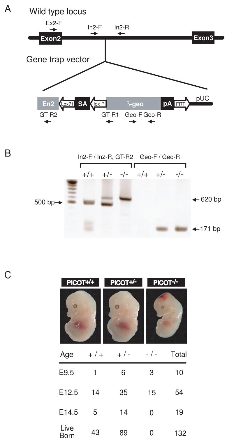
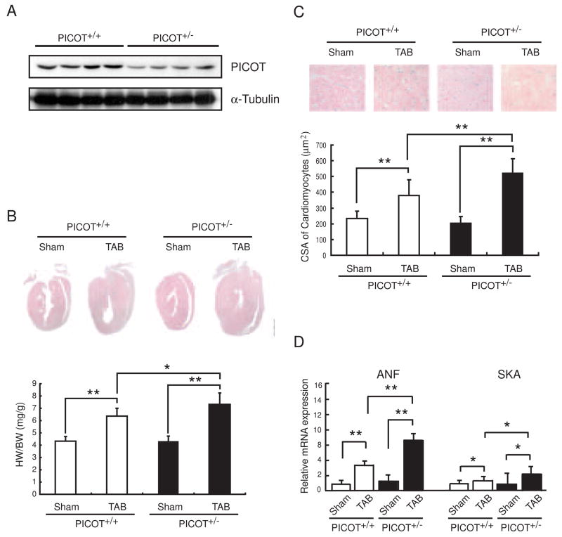
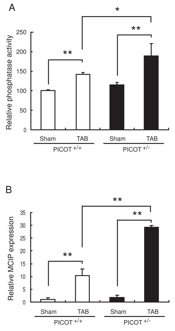
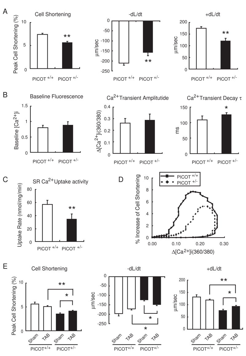
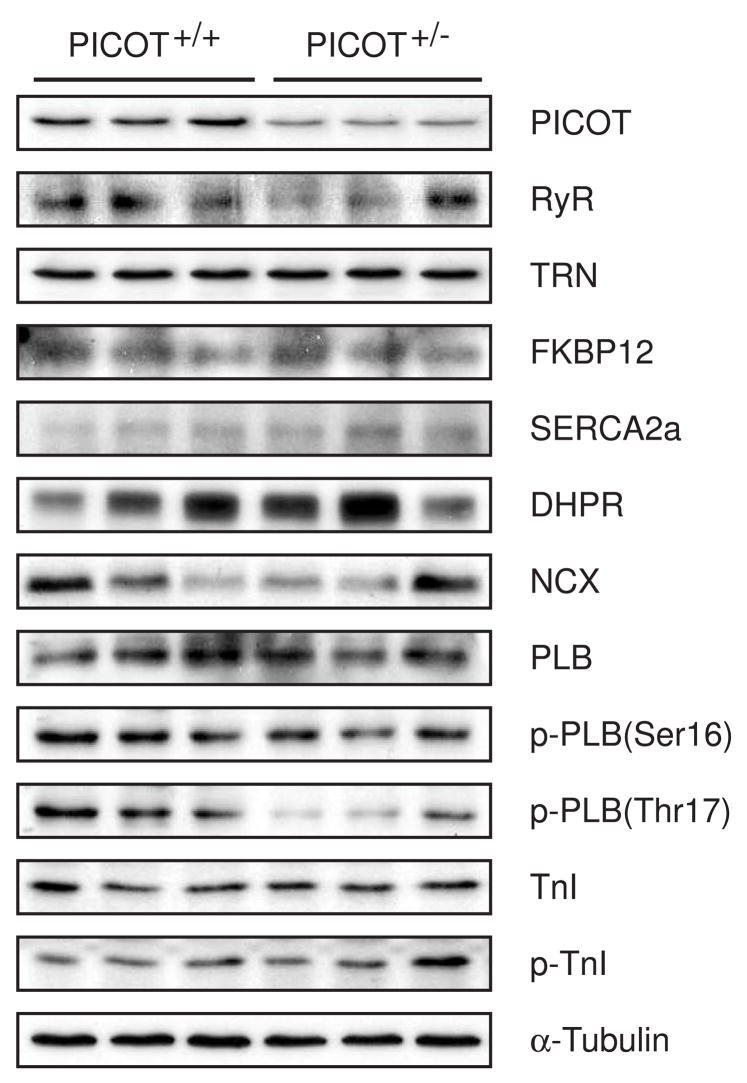
PICOT (PKC-interacting cousin of thioredoxin) was previously shown to inhibit the development of cardiac hypertrophy, concomitant with an increase in cardiomyocyte contractility. To explore the physiological function of PICOT in the hearts, we generated a PICOT-deficient mouse line by using a gene trap approach. PICOT(-/-) mice were embryonic lethal indicating that PICOT plays an essential role during embryogenesis, whereas PICOT(+/-) mice were viable with no apparent morphological defects. The PICOT protein levels were reduced by about 50% in the hearts of PICOT(+/-) mice. Significantly exacerbated cardiac hypertrophy was induced by pressure overload in PICOT(+/-) mice relative to that seen in wild type littermates. In line with this observation, calcineurin-NFAT signaling was greatly enhanced by pressure overload in the hearts of PICOT(+/-) mice. Cardiomyocytes from PICOT(+/-) mice exhibited significantly reduced contractility, which may be due in part to hypophosphorylation of phospholamban and reduced SERCA activity. These data indicate that the precise PICOT protein level significantly affects the process of cardiac hypertrophy and cardiomyocyte contractility. We suggest that PICOT plays as a critical negative regulator of cardiac hypertrophy and a positive inotropic regulator.


Figures

Figure 1
Figure 1. Targeted disruption of the PICOT gene
(A) Embryonic stem cell line RRF094 contains the gene trap vector pGT0 inserted within the second intron of PICOT. The PICOT locus and pGT0 insertion are shown. The vector pGT0 contains a part of the mouse engrailed 2 intron 1 (En2), a splice acceptor of mouse En2 exon 2 (SA), a β-galactosidase/neomycin resistance fusion gene (β-geo), and a SV40 polyadenylation sequence (pA). Arrows indicate PCR primer binding sites for genomic PCR and genotyping. (B) Representative genotypic analysis of littermates harboring the WT (+) or deleted allele (−) of the PICOT gene from a PICOT+/− intercrosses. Genotyping was performed by PCR using primers In2-F and In2-R for the wild-type (500 bp), and In2-F and GT-R2 for the deleted allele (620 bp). The presence of gene trap vector was also confirmed by PCR using primer pairs Geo-F and Geo-R (171 bp). (C) Photographs of littermates (E12.5) from PICOT+/− intercrosses are shown. The genotype of the littermates was analyzed by PCR.
Figure 2
Figure 2. Enhanced hypertrophy in PICOT+/− mice
(A) Adult heart lysates from PICOT+/+ and PICOT+/− littermates were separated by SDS-PAGE and subjected to Western blot analysis with anti-PICOT antibody. (B) Representative longitudinal sections of PICOT+/+ and PICOT+/− hearts, sham operated (Sham) or transverse aortic banded for one week (TAB) are shown. The heart weight (HW) to body weight (BW) ratio is shown. n=6 per group. (C) Higher magnification photographs of the heart sections are also shown. Cell surface area (CSA) of individual cardiomyocyte was measured using the AnalSIS image analysis program. n=3 per group; about 100 cells were analyzed from each heart. (D) Expression levels of hypertrophic marker genes, ANF and SKA, were determined by quantitative RT-PCR. All the data are represented as mean ± SD. *P<0.05, **P<0.01.
Figure 3
Figure 3
Enhanced calcineurin-NFAT signaling in PICOT+/− mice. (A) Calcineurin phosphatase activity was determined. (B) Expression level of a transcriptional target of NFAT, MCIP, was determined by quantitative RT-PCR. All the data are represented as mean ± SD. *P<0.05, **P<0.01.
Figure 4
Figure 4. Cardiomyocyte contractility and Ca2+ transient in PICOT+/− mice
(A) Adult cardiomyocytes were isolated from PICOT+/+ and PICOT+/− littermates and their contractile parameters were determined. Peak cell shortening, percentage of shortened cell length; −dL/dt, maximal velocities of cell shortening; +dL/dt, maximal velocities of cell relengthening. (B) Average parameters of Ca2+ transient properties were determined using fura2/acetoxymethyl ester. (C) SR Ca2+ uptake rates were measured in the presence of 0.2 mM CaCl2. (D) A plot of hysteresis loop of cell shortening against cytosolic Ca2+ content is shown. (E) Adult cardiomyocytes were isolated from PICOT+/+ and PICOT+/− littermates which were sham operated (Sham) or transverse aortic banded (TAB) for 1 week and their contractile parameters were determined. Approximately 140 cells were chosen for the contractility and Ca2+ transient measurements from seven individual hearts per group. All the data are represented as mean ± SD. For panels A–C, *P<0.05 vs PICOT+/+ and **P<0.01 vs PICOT+/+. For panel E, *P<0.05 and **P<0.01.
Figure 5
Figure 5. Western blot analysis of heart lysates
Heart lysates from PICOT+/− and PICOT+/− littermates were separated by SDS-PAGE, transferred to PVDF filter, and probed with antibodies against PICOT, PKC-interacting cousin of Thioredoxin; RyR, ryanodine receptor; TRN, triadin; FKBP12, FK506 binding protein 12; SERCA2a, sarcomeric reticular Ca2+ ATPase 2a; DHPR, dihydropyridine receptor; NCX, Na2+-Ca2+ exchanger; PLB, phospholamban; p-PLB, phosphorylated PLB; TnI, troponin I; p-TnI, phosphorylated TnI. α-tubulin was used a loading control.

References

    1. Heineke J, Molkentin JD. Regulation of cardiac hypertrophy by intracellular signalling pathways. Nature reviews. 2006 Aug;7(8):589–600. - PubMed
    1. Zimmer HG, Gerdes AM, Lortet S, Mall G. Changes in heart function and cardiac cell size in rats with chronic myocardial infarction. Journal of molecular and cellular cardiology. 1990 Nov;22(11):1231–43. - PubMed
    1. Rockman HA, Ross RS, Harris AN, Knowlton KU, Steinhelper ME, Field LJ, et al. Segregation of atrial-specific and inducible expression of an atrial natriuretic factor transgene in an in vivo murine model of cardiac hypertrophy. Proceedings of the National Academy of Sciences of the United States of America. 1991 Sep 15;88(18):8277–81. - PMC - PubMed
    1. Lips DJ, deWindt LJ, van Kraaij DJ, Doevendans PA. Molecular determinants of myocardial hypertrophy and failure: alternative pathways for beneficial and maladaptive hypertrophy. European heart journal. 2003 May;24(10):883–96. - PubMed
    1. Berenji K, Drazner MH, Rothermel BA, Hill JA. Does load-induced ventricular hypertrophy progress to systolic heart failure? American journal of physiology. 2005 Jul;289(1):H8–H16. - PubMed

Publication types

MeSH terms

Substances