. 2008 Dec 15;45(12):1682-94.
doi: 10.1016/j.freeradbiomed.2008.09.009. Epub 2008 Sep 23.

Identification and characterization of VPO1, a new animal heme-containing peroxidase

Affiliations

Identification and characterization of VPO1, a new animal heme-containing peroxidase

Guangjie Cheng et al. Free Radic Biol Med. .

Abstract

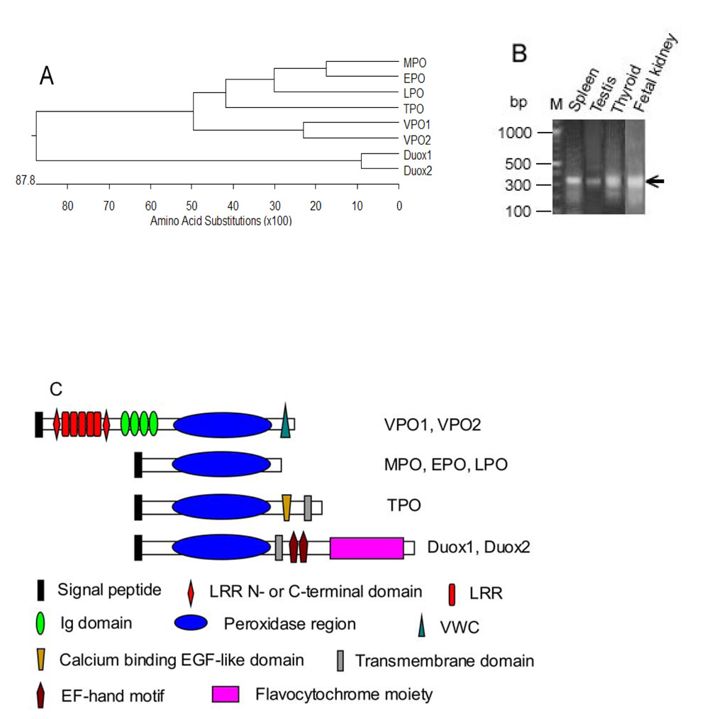
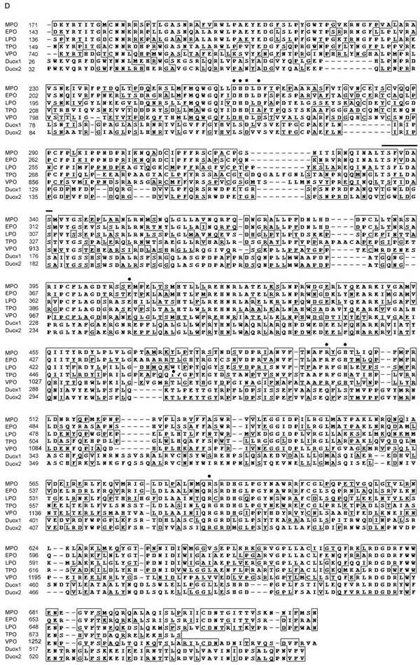
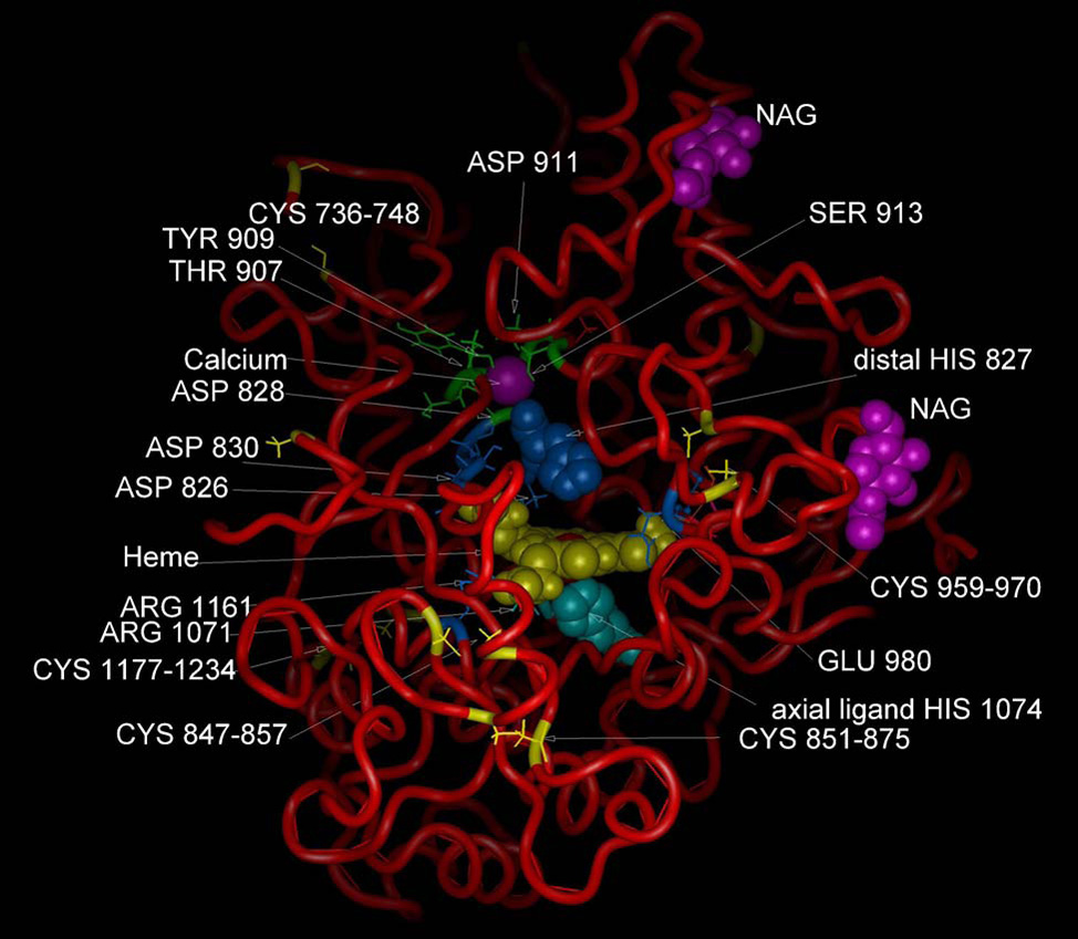
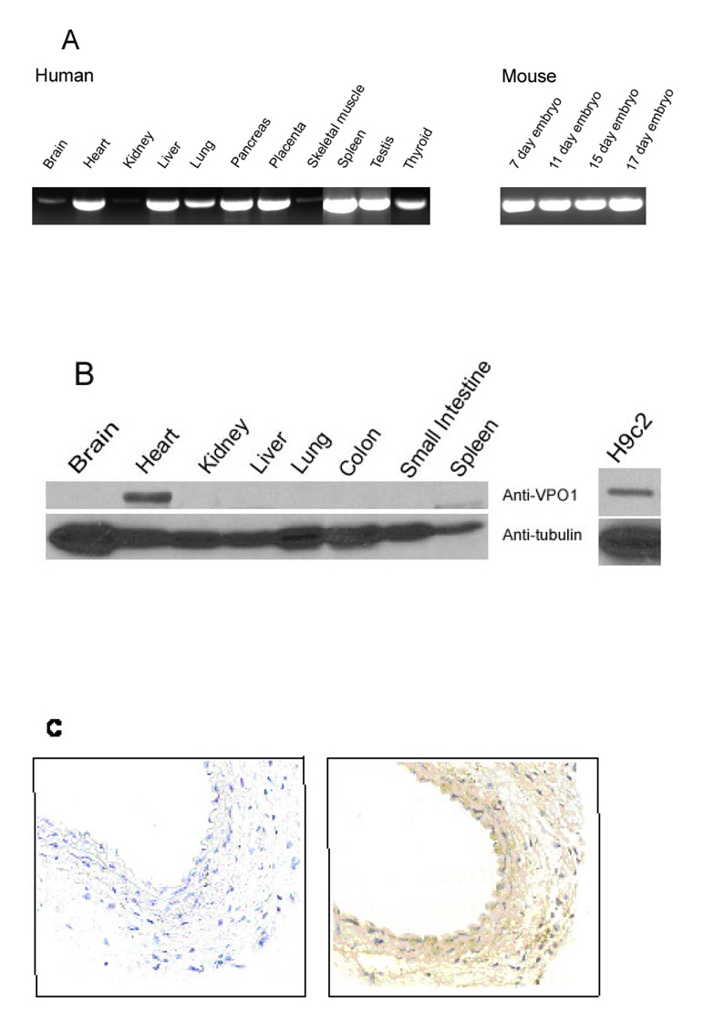
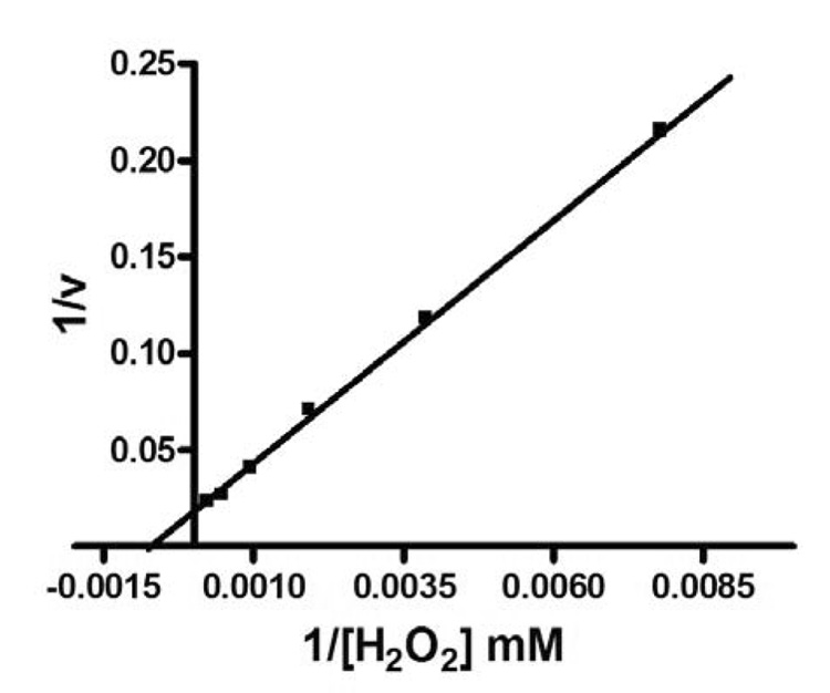
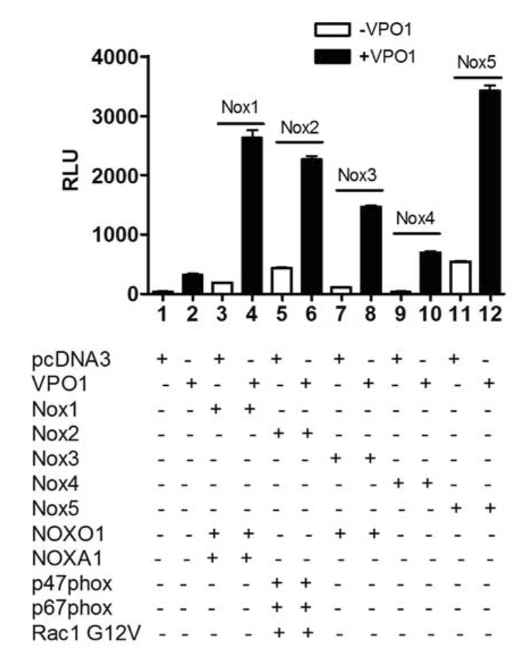
Animal heme-containing peroxidases play roles in innate immunity, hormone biosynthesis, and the pathogenesis of inflammatory diseases. Using the peroxidase-like domain of Duox1 as a query, we carried out homology searching of the National Center for Biotechnology Information database. Two novel heme-containing peroxidases were identified in humans and mice. One, termed VPO1 for vascular peroxidase 1, exhibits its highest tissue expression in heart and vascular wall. A second, VPO2, present in humans but not in mice, is 63% identical to VPO1 and is highly expressed in heart. The peroxidase homology region of VPO1 shows 42% identity to myeloperoxidase and 57% identity to the insect peroxidase peroxidasin. A molecular model of the VPO1 peroxidase region reveals a structure very similar to that of known peroxidases, including a conserved heme binding cavity, critical catalytic residues, and a calcium binding site. The absorbance spectra of VPO1 are similar to those of lactoperoxidase, and covalent attachment of the heme to VPO1 protein was demonstrated by chemiluminescent heme staining. VPO1 purified from heart or expressed in HEK cells is catalytically active, with a K(m) for H(2)O(2) of 1.5 mM. When co-expressed in cells, VPO1 can use H(2)O(2) produced by NADPH oxidase enzymes. VPO1 is likely to carry out peroxidative reactions previously attributed exclusively to myeloperoxidase in the vascular system.


Figures

Fig. 1
Fig. 1
Identification of VPO1. A. Shown is a dendrogram showing the similarity based on sequence identity among the peroxidase domains of human heme-containing peroxidases. B. Shown are results from 5’-RACE of VPO1 from four human tissues. 5’-RACE was carried out as described in Materials and Methods. The 350 bp bands (arrow) from the four tissues were extracted from the gel and verified by DNA sequencing. M is DNA size marker and its size is shown on left. C. Shown are predicted domain or motif structures of VPO1, VPO2 and other heme-containing peroxidases. D. Alignment of amino acid sequences of the peroxidase domains of the human heme-containing peroxidases. Filled circles indicate conserved residues that in MPO are predicted to form heme binding cavity[24]. The residues of axial and distal histidine are at 1074 and 827, respectively. The highly conserved sites for binding to calcium are indicated by filled square (Asp828) and the horizontal superior line. All these conserved residues are shown in homology model of VPO1 (Fig.2).
Fig. 1
Fig. 1
Identification of VPO1. A. Shown is a dendrogram showing the similarity based on sequence identity among the peroxidase domains of human heme-containing peroxidases. B. Shown are results from 5’-RACE of VPO1 from four human tissues. 5’-RACE was carried out as described in Materials and Methods. The 350 bp bands (arrow) from the four tissues were extracted from the gel and verified by DNA sequencing. M is DNA size marker and its size is shown on left. C. Shown are predicted domain or motif structures of VPO1, VPO2 and other heme-containing peroxidases. D. Alignment of amino acid sequences of the peroxidase domains of the human heme-containing peroxidases. Filled circles indicate conserved residues that in MPO are predicted to form heme binding cavity[24]. The residues of axial and distal histidine are at 1074 and 827, respectively. The highly conserved sites for binding to calcium are indicated by filled square (Asp828) and the horizontal superior line. All these conserved residues are shown in homology model of VPO1 (Fig.2).
Fig. 2
Fig. 2
Predicted molecular structure of VPO1. A structural homology model was constructed as described in Materials and Methods, and utilized LPO, MPO and PGS as a basis set. Heme is indicated in yellow. Calcium ion is shown in purple and predicted residues binding to calcium are Asp828, Thr907 Tyr909 and Asp911. There are two putative N-acetylglucosamine (NAG) sugar moieties shown. Residues comprising the conserved heme-binding cavity are shown. These include conserved axial and distal histidines (1074 and 827, respectively), residues cross-linking to the vinyl groups of heme (Asp826 and Glu980) and residues interacting with the heme propionate group (Asp830, Arg1071,and Arg1161) [24]. The highly conserved Cys-Cys disulfides are also indicated.
Fig. 3
Fig. 3
Tissue distribution of VPO1. A. RT-PCR was used to detect VPO1 in a variety of tissues as indicated using human- and mouse-specific PCR primers as described in Materials and Methods. The identity of the PCR fragment from human heart and mouse 17 day embryo was confirmed by DNA sequencing. B. A Western blotting was carried out as described in Materials and Methods using Protein-G purified anti-VPO1 polyclonal antibody in mouse (left); and in the rat myocardium cell line H9c2 (right). The lower panel uses anti-tubulin antibody as loading control. C. Immunohistochemistry of mouse carotid artery. Immunohistochemistry was performed as described in Materials and Methods. In the left panel primary antibody is omitted. The primary antibody used in the right panel was the same anti-VPO1 antibody as in B.
Fig. 4
Fig. 4
Reconstitution of VPO1 activity. A. Effect of NaBu and hematin on cellular peroxidase activity. Empty vector (1) or expression vector encoding VPO1 (2) was transfected into HEK 293H cells as described in Materials and Methods. The cells were cultured in 5 mM of NaBu and 1 µg/ml of hematin at 37%, 5% CO2 for 24 hrs before harvest. The TMB oxidation was carried out using lyzed cells. Error bars show the range of three independent experiments. Inset shows a Western blot indicating VPO1 expression in one of the experiment. B. VPO1-stably expressing cells were incubated with 5 mM NaBu and 1 µg/ ml hematin for indicated time. Western blotting was carried out to determine the VPO1 expression levels using anti-VPO1 antibody while the same membrane was blotted with anti-tubulin antibody as a loading control. C. Optimization of NaBu concentration. Cells were grown to 80% confluence, and 1 µg/ml hematin plus the indicated concentration of NaBu were added to the media and cells were cultured for an additional 24 hrs. TMB oxidation was carried out as described in A. GJ-3 and GJ-4 are two cell colonies that stably-expressing VPO1 and are derived from HK293H cells as described in Materials and Methods. D. Peroxidase activity in living cells. Luminol-based chemiluminescence was measured in living cells as described in Materials and Methods at the indicated concentrations of exogenously added H2O2. Data are representative of three independent experiments. RLU refers to relative light units. E. Inhibition of VPO1-dependent peroxidation of TMB by ABH. HEK293H and GJ-4 cells were incubated with 5 mM NaBu and 1 µg/ ml hematin for 24 hrs before harvesting. TMB oxidation was measured as described in A except that the indicated concentration of ABH was added to the incubation.
Fig. 5
Fig. 5
UV-visible absorbance spectra of human partially purified full-length VPO1. A. Spectra of expressed, partially purified VPO1 are shown, including oxidized VPO1, oxidized VPO1 in pyridine (pyr), reduced VPO1 in pyridine, and oxidized VPO1 in 88% formic acid. B. Reduced spectra of pyridine hemochrome. Reduced spectra of pyridine hemochrome were obtained by addition of pyridine to 2.4 M and a few crystals of sodium dithionite. The reduced spectrum showed a split Soret band with features at 393 and 413 nm. The presence of the symmetrical Soret peak of VPO1 in above conditions provides evidence for a native environment for the heme. C. Spectra from 450 nm to 700 nm of B.
Fig. 6
Fig. 6
VPO1 covalently binds to heme. The experimental procedures are described as in Materials and Methods. The blot in the upper panel was developed with Pierce’s chemiluminescent substrate and chemiluminescent signal was detected by exposure to X-ray film. After extensive washing, the same blot was stained with Coomassie’s Blue and destained using standard procedures (lower panel). 1. myoglobin; 2. cytochrome c; 3. VPO1. Molecular size scale is shown on the left.
Fig. 7
Fig. 7
Lineweaver-Burk plot of TMB oxidation by VPO1. Inverse rate data were plotted as a function of the inverse of H2O2 concentration. The Km for H2O2 using TMB as a substrate was determined as described in Materials and Methods. Data are the average of three independent experiments.
Fig. 8
Fig. 8
VPO1 utilizes H2O2 generated by Nox enzymes. Plasmids encoding the indicated Noxes or Nox regulatory proteins were transfected into HEK293H cells as described in Materials and Methods. After 24 hr, NaBu and hematin were added as in Fig. 4 and cells were allowed to continue in culture for an additional 24 hrs. Luminol-based chemiluminescence was then detected in the absence of added H2O2.The data are representative of three independent experiments. For the Nox2 group, the cells were treated with 200 nM phorbol 12-myristate 13-acetate at 37°C for 10 min prior to the assay.

References

    1. Klebanoff SJ. Myeloperoxidase: friend and foe. J Leukoc Biol. 2005;77:598–625. - PubMed
    1. Edens WA, Sharling L, Cheng G, Shapira R, Kinkade JM, Lee T, Edens HA, Tang X, Sullards C, Flaherty DB, Benian GM, Lambeth JD. Tyrosine cross-linking of extracellular matrix is catalyzed by Duox, a multidomain oxidase/peroxidase with homology to the phagocyte oxidase subunit gp91phox. J Cell Biol. 2001;154:879–891. - PMC - PubMed
    1. Salmon SE, Cline MJ, Schultz J, Lehrer RI. Myeloperoxidase deficiency. Immunologic study of a genetic leukocyte defect. The New England journal of medicine. 1970;282:250–253. - PubMed
    1. Lehrer RI, Cline MJ. Leukocyte myeloperoxidase deficiency and disseminated candidiasis: the role of myeloperoxidase in resistance to Candida infection. J Clin Invest. 1969;48:1478–1488. - PMC - PubMed
    1. Mansson-Rahemtulla B, Rahemtulla F, Baldone DC, Pruitt KM, Hjerpe A. Purification and characterization of human salivary peroxidase. Biochemistry. 1988;27:233–239. - PubMed

Publication types

MeSH terms