. 2008 Dec 15;480(2):85-94.
doi: 10.1016/j.abb.2008.09.016. Epub 2008 Oct 7.

4-Hydroxynonenal induces p53-mediated apoptosis in retinal pigment epithelial cells

Affiliations

4-Hydroxynonenal induces p53-mediated apoptosis in retinal pigment epithelial cells

Abha Sharma et al. Arch Biochem Biophys. .

Abstract

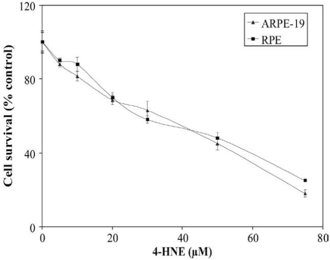
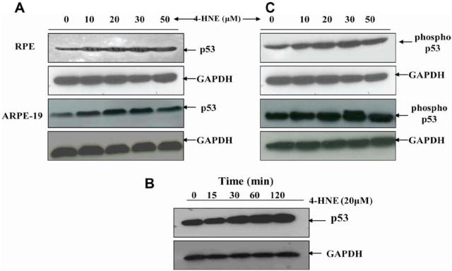
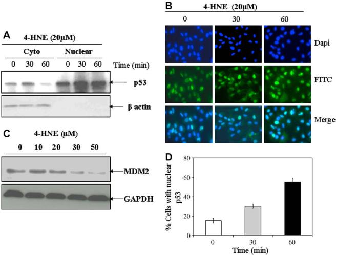
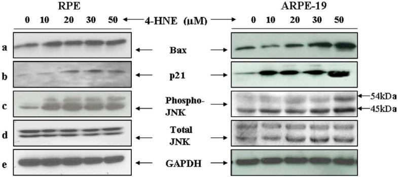
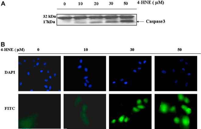
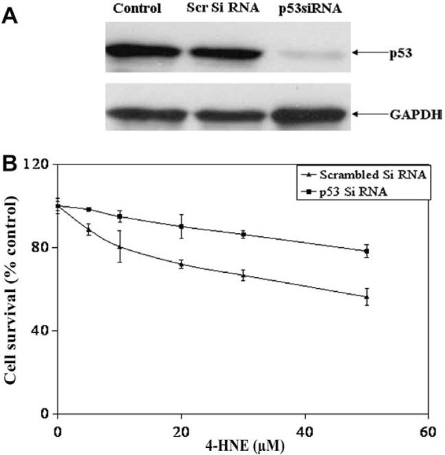
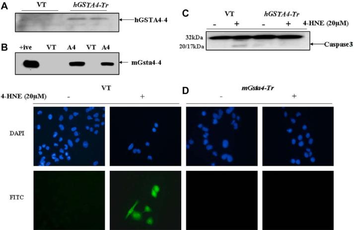
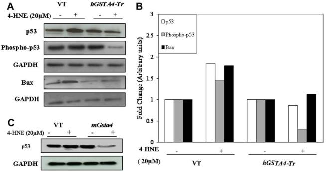
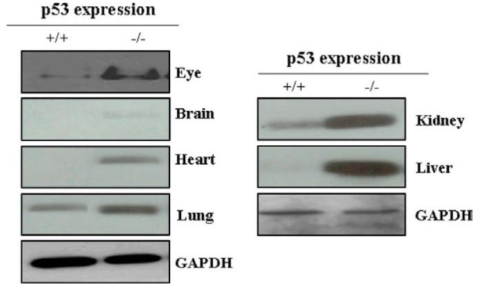
4-Hydroxynonenal (4-HNE) has been suggested to be involved in stress-induced signaling for apoptosis. In present studies, we have examined the effects of 4-HNE on the intrinsic apoptotic pathway associated with p53 in human retinal pigment epithelial (RPE and ARPE-19) cells. Our results show that 4-HNE causes induction, phosphorylation, and nuclear accumulation of p53 which is accompanied with down regulation of MDM2, activation of the pro-apoptotic p53 target genes viz. p21 and Bax, JNK, caspase3, and onset of apoptosis in treated RPE cells. Reduced expression of p53 by an efficient silencing of the p53 gene resulted in a significant resistance of these cells to 4-HNE-induced cell death. The effects of 4-HNE on the expression and functions of p53 are blocked in GSTA4-4 over expressing cells indicating that 4-HNE-induced, p53-mediated signaling for apoptosis is regulated by GSTs. Our results also show that the induction of p53 in tissues of mGsta4 (-/-) mice correlate with elevated levels of 4-HNE due to its impaired metabolism. Together, these studies suggest that 4-HNE is involved in p53-mediated signaling in in vitro cell cultures as well as in vivo that can be regulated by GSTs.


Figures

Fig. 1
Fig. 1
Cytotoxicity of 4-HNE to RPE and ARPE-19 cells: Cells (2 × 10⁴) were plated in 96-well plates in complete growth medium for 24 h to allow for complete attachment to the culture plates. Next day, cells were incubated for 18 h in fresh serumfree medium to avoid any interaction between serum proteins and 4-HNE and solutions containing various amounts of 4-HNE prepared in PBS were added to achieve the final concentrations of 0-75 μM 4-HNE. Eight replicate wells were used for each concentration of 4-HNE used in these studies and the plates were incubated at 37 °C for 2 h. The MTT assay was performed as described in Materials and methods section. The OD562 values of samples subtracted from those of respective blanks (no cells) were normalized with control values. Results presented are percent cell survital in 4-HNE treated groups with respect to control cells (mean ± SD; n = 8).
Fig. 2
Fig. 2
Effect of 4-HNE on induction and phosphorylation of p53 in RPE and ARPE-19 cells: Western blot analyses of 4-HNE treated RPE and ARPE-19 cells showing the activation of p53 with dose (A) and time (B), and p53 phosphorylation (C). For (A) and (C) cells were treated with 0-50 μM 4-HNE in the medium for 2 h. To assess the time dependent activation of p53 (B), RPE cells (4 × 10⁵) were treated with 20 μM 4-HNE and incubated for 0, 15, 30, 60, and 120 min. Cells were harvested after completion of incubation and extracted in RIPA lysis buffer as described in the Methods section. Cell extracts (50 μg protein) were resolved on 4-20% SDS-PAGE and immunoblotted using anti-p53 (A), (B) and anti-phospho p53 (serine-15) (C) antibodies, respectively as the primary antibodies. GAPDH was used as the loading control. Blots were developed with West Pico-chemiluminescence reagent (Pierce). Representative blot from three different experiments yielding similar results has been presented.
Fig. 3
Fig. 3
4-HNE-induced nuclear accumulation of p53 and degradation of MDM2 in RPE cells: RPE cells (2 × 10⁶ cells in 100 mm petri dishes and 1 × 10⁴ cells/chamber) were treated with 20 μM 4-HNE for different time intervals (0, 30, and 60 min). (A) Cells were harvested from the petri dishes and their cytoplasmic and nuclear extracts were prepared by using Imgenex cell processing kit according to the manufacturer's instructions. Western blot analyses for the expression of p53 in cytoplasmic and nuclear extracts (50 μg protein) were carried out using anti-p53 antibodies. Western blot presented show a time dependent decrease of p53 in cytoplasmic fractions with corresponding increase of p53 in the nuclear fractions. (B) 4-HNE treated cells in chamber slides were fixed in 4% paraformaldehyde, blocked with 1% goat serum and incubated with anti-p53 antibodies overnight at 4 °C. The cells were washed with PBS, incubated with FITC-conjugated secondary antibodies for 2 h, mounted with Vectashield mounting medium containing DAPI nuclear stain and the slides were viewed under Olympus fluorescence microscope. Photographs were taken with 40× objective. Photomicrographs show an enhanced nuclear accumulation of p53 with time as judged by the increase in green fluorescent stain in the nucleus. (C) Western blot analysis showing a gradual decrease of MDM2 levels in RPE cells treated with 0-50 μM 4-HNE for 2 h. Blots were probed with anti-MDM2 polyclonal antibodies. (D) Bar chart showing the percentage of cells with 4-HNE (20 μM) induced nuclear accumulation of p53 at different time points (mean ± SD, n = 3). (For interpretation of color mentioned in this figure the reader is referred to the web version of the article.)
Fig. 4
Fig. 4
4-HNE-induced activation of Bax, p21, JNK, and p-JNK in RPE and ARPE-19 cells: The expression of Bax (a) p21 (b) p-JNK (c), and JNK (d) in RPE and ARPE-19 cells exposed to different concentrations of 4-HNE. RPE and ARPE-19 cells were treated with 4-HNE (0-50 lM) for 2 h in serum-free medium. Cell extracts (50 μg protein) were resolved on 4-20% SDS-PAGE and immunoblotted. The blots were probed with anti-Bax, anti-p21, anti-JNK, and anti-p-JNK antibodies, respectively. GAPDH (e) was used as the loading control. Blots were developed with West Pico-chemi luminescence reagent (Pierce).
Fig. 5
Fig. 5
Effect of 4-HNE on caspase3 in RPE and ARPE-19 cells: (A) Cell extracts (50 μg protein) from RPE cells treated with 4-HNE (0-50 μM) for 2 h were resolved on 4-20% SDS-PAGE and immunoblotted using the anti-caspase3 antibody as the primary antibody. Activation of caspase3 was monitored by the appearance of the 20/17 kDa bands. The blot was developed using West Pico-chemiluminescence reagent (Pierce). (B) In situ analysis of activated caspase3 in ARPE-19 cells. About 2 × 10⁴ cells were grown in chamber slides and treated with 0, 10, 30, 50 μM 4-HNE for 2h. The activation of caspase3 in these cells was examined by staining with 10 μM CaspACE™ FITC-VAD-FMKin situ marker following the manufacturer's instructions. The slides were mounted with Vectashield DAPI mounting medium and observed with a fluorescence microscope (Olympus) using the standard filter sets for DAPI and FITC. Appropriately marked different panels show blue DAPI-stained and green FITC-stained cells in the figure. The photographs were taken at 400× magnification. (For interpretation of color mentioned in this figure the reader is referred to the web version of the article.)
Fig. 6
Fig. 6
Silencing of p53 protects RPE cells from 4-HNE induced cytotoxicity: (A) RPE cells (1 × 10⁵) were transfected with si RNA of p53 and scrambled siRNA as described in Methods section. The depletion of p53 in cells was confirmed by Western blot analysis. (B) P53 depleted and p53 expressing control cells were treated with 0-50 μM of 4-HNE for 2 h and assayed for cytotoxicity by MTT assay. The plot shows the percent cell survival (mean ± SD, n = 4) at different concentrations of 4-HNE.
Fig. 7
Fig. 7
Effect of GSTA4 transfection on 4-HNE-induced caspase3 activation and apoptosis: (A) Expression of hGSTA4-4 and (B) mGsta4-4 in the transfected RPE and ARPE-19 cells, respectively. Aliquots (50 μg) of 28,000 g supernatant fraction of homogenates of the vector (VT) and hGSTA4-transfected (hGSTA4-Tr) RPE cells and mGsta4-transfected ARPE-19 cells were subjected to SDS-PAGE in 4-20% gels. Western blot analyses were performed using the polyclonal primary antibody against recombinant hGSTA4-4 and mGsta4-4. The blot was developed using chemiluminescence (Supersignal West Pico, Pierce) reagents. (C) Western blot analysis for caspase3 activation in the VT and hGSTA4-Tr RPE cells treated with 20 μM 4-HNE for 1 h. Cell extracts (50 μg protein) were resolved on 4-20% SDS-PAGE and immunoblotted using the anti-caspase3 antibody as the primary antibody. Activation of caspase3 was monitored by the appearance of 20/17 kDa band in the VT transfected, 20 μM 4-HNE-treated RPE cells only. The blots were developed using chemiluminescence (Supersignal West Pico, Pierce) reagents. (D) In situ analysis of activated caspase3 in VT and mGsta4-Tr RPE cells. 1 × 10⁵ RPE cells were treated with 20 μM 4-HNE for 1 h. The activation of caspase3 in these cells was examined by staining with 10 μM CaspACE™ FITC-VAD-FMK in situ marker following the manufacturer's instructions. The slides were mounted with Vectashield DAPI mounting medium and observed with a fluorescence microscope (Olympus) using the standard filter sets for DAPI and FITC. Different panels show blue DAPI-stained and green FITC-stained cells as marked in the figure. Green FITC-labeled VT cells show a significant increase in the level of caspase3 activation after 4-HNE treatment. (For interpretation of color mentioned in this figure the reader is referred to the web version of the article.)
Fig. 8
Fig. 8
Effect of 4-HNE on the expression of p53, phospho p53, and Bax in GSTA4-4 over-expressing RPE cells: (A) Expression of p53, phospho p53, and Bax, in VT and hGSTA4-Tr RPE cells treated with 4-HNE (20 μM) for 1 h was monitored by Western blot analysis. Cell extracts (50 μg protein) were resolved on 4-20% SDS-PAGE and immunoblotted using the anti-p53, anti-phospho p53, and anti-Bax, respectively as the primary antibodies. GAPDH was used as the loading control. The blot was developed using chemiluminescence (Supersignal West Pico, Pierce) reagents. (B) A bar graph showing densitometric analysis of bands (using Kodak 1D image analysis software) of p53, phospho p53, and Bax, respectively. (C) Effect of 4-HNE on p53 expression in mGsta4-4 overexpressing ARPE-19 cells.
Fig. 9
Fig. 9
Expression of p53 in different tissues of mGSTA4 (+/+) and (-/-) mice: Mice eye, brain, heart, lung, kidney, and liver were homogenized in lysis buffer containing 50 mM Tris-HCl (pH 7.5), 1 mM sodium orthovanadate, 50 mM NaF, PMSF (20 μg/ml), 4 mM EDTA, 1 mM EGTA, 1% NP-40, 150 mM sodium chloride, 100 μM leupeptin, 0.07 μg/ml pepstatin, 10 μg/ml soybean trypsin inhibitor, 1:100 protease inhibitor cocktail and centrifuged at 14000 rpm. Supernatants containing 50 μg tissue proteins were loaded on 4-20% SDS-PAGE and p53 was assayed by Western blot analysis using anti-p53 antibody as the primary antibody. GAPDH was used as a loading control. Blots were developed with West Pico-chemiluminescence reagent (Pierce).

References

    1. Esterbauer H, Schaur RJ, Zollner H. Free Radic. Biol. Med. 1991;11:81–128. - PubMed
    1. Awasthi YC, Sharma R, Cheng JZ, Yang Y, Sharma A, Singhal SS, Awasthi S. Mol. Aspects Med. 2003;24:219–230. - PubMed
    1. Dianzani MU. Mol. Aspects Med. 2003;24:263–272. - PubMed
    1. Levine AJ. Cell. 1997;88:323–331. - PubMed
    1. Gottlieb TM, Oren M. Semin. Cancer Biol. 1998;8:359–368. - PubMed

Publication types