Review
. 2008 Dec;1782(12):691-9.
doi: 10.1016/j.bbadis.2008.10.002. Epub 2008 Oct 10.

Autophagy and the ubiquitin-proteasome system: collaborators in neuroprotection

Affiliations
Review

Autophagy and the ubiquitin-proteasome system: collaborators in neuroprotection

Natalia B Nedelsky et al. Biochim Biophys Acta. 2008 Dec.

Abstract

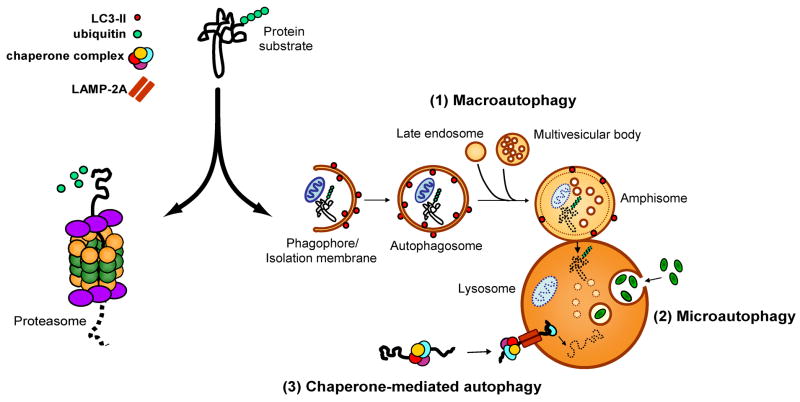
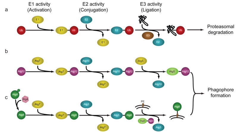
Protein degradation is an essential cellular function that, when dysregulated or impaired, can lead to a wide variety of disease states. The two major intracellular protein degradation systems are the ubiquitin-proteasome system (UPS) and autophagy, a catabolic process that involves delivery of cellular components to the lysosome for degradation. While the UPS has garnered much attention as it relates to neurodegenerative disease, important links between autophagy and neurodegeneration have also become evident. Furthermore, recent studies have revealed interaction between the UPS and autophagy, suggesting a coordinated and complementary relationship between these degradation systems that becomes critical in times of cellular stress. Here we describe autophagy and review evidence implicating this system as an important player in the pathogenesis of neurodegenerative disease. We discuss the role of autophagy in neurodegeneration and review its neuroprotective functions as revealed by experimental manipulation in disease models. Finally, we explore potential parallels and connections between autophagy and the UPS, highlighting their collaborative roles in protecting against neurodegenerative disease.


Figures

Figure 1
Figure 1
The UPS and the autophagy-lysosomal systems are the two main protein degradation systems in the cell. Protein that are tagged with polyubiquitin chains are generally considered to be substrates for the UPS, which feeds unfolded proteins through the barrel of the 26S proteasome and generates small digested peptides. Recent evidence suggests that some ubiquitinated substrates can also be degraded via the autophagy-lysosomal system. This system is comprised of (1) chaperone-mediated autophagy (CMA), in which soluble substrates associated with a specific chaperone complex are translocated into the lysosome through the LAMP-2A lysosomal receptor, (2) microautophagy, in which small volumes of cytosol are directly engulfed by lysosomes, and (3) macroautophagy, in which cytosolic components are engulfed and delivered to the lysosome in bulk. Macroautophagy involves a series of maturation steps: first, a portion of cytoplasm is surrounded by an expanding elongation membrane or phagophore. The phagophore seals to form an autophagosome, which in mammals fuses with late endosomes and multivesicular bodies to form an amphisome. The amphisome then fuses with a lysosome to form an autolysosome, in which cytosolic cargo is degraded by lysosomal hydrolases. LC3-II is a protein that associates with the inner and outer surfaces of autophagic membranes and provides a histological marker of autophagic vacuoles.
Figure 2
Figure 2
Assembly and elongation of autophagic membranes are accomplished via sequential action of UPS-like E1-E2-E3 cascades. In each case, an E1 enzyme activates a ubiquitin-like protein (UBL) such as ubiquitin, Atg12, or Atg8. The UBL is then transferred to an E2 conjugating enzyme, followed by an association with an E3 ligase that promotes association of the UBL and its target. (a) In the UPS, ubiquitination of substrates is accomplished by an E1-activating enzyme, E2-conjugating enzyme, and an E3-ligase. (b) In the first arm of the Atg conjugation pathway, Atg12 associates with the E1-like Atg7, is transferred to the E2-like Atg10, and is subsequently conjugated to Atg5. No E3-like protein has been identified in this pathway. (c) In the second arm of the Atg conjugation pathway, Atg8 associates with the E1-like Atg7, is transferred to the E2-like Atg3, and is conjugated to PE via the E3-like action of the Atg12-Atg5 complex. Adapted from [101] with permission.
Figure 3
Figure 3
Proteasome impairment leads to upregulation of autophagic activity. (a) HeLa cells that stably express the UPS reporter UbG76V-GFP were treated with the irreversible proteasome inhibitor epoximicin for 72 hours and monitored for cell death. Increasing levels of the GFP substrate indicate impaired UPS function. Note the 24 hours time point used in (b)–(g) is within the window during which proteasome function is impaired, but the cells remain viable. (b–c) Images of LC3 staining (red) and DAPI (blue) show accumulation of LC3 puncta in epoximicin-treated cells. (d–e) Transmission electron microscopy images of cells reveal autophagic structures and prominent vacuolization in epoximicin-treated cells. (f–g) Increased magnification of structures in (e) reveal multi-membraned structures consistent with autophagic activity.
Figure 4
Figure 4
A Drosophila model of proteasome impairment is modified by manipulation of autophagic activity. (a–b) The temperature-sensitive DTS7 mutant shows a normal eye phenotype at the permissive temperature of 22°C and a significant degenerative phenotype at the restrictive temperature of 28°C. (c) RNAi knockdown of the autophagy gene atg12 results in an enhancement of the DTS7 degenerative phenotype, suggesting that the autophagic activity that is induced in response to proteasome impairment is compensatory. (d) Treatment of DTS7 flies with rapamycin suppresses the degenerative phenotype, demonstrating that induction of autophagy can compensate for impaired proteasome function. Adapted from [50].

Similar articles

Cited by

References

    1. Wheatley DN, Inglis MS. An intracellular perfusion system linking pools and protein synthesis. J Theor Biol. 1980;83:437–445. - PubMed
    1. Vabulas RM, Hartl FU. Protein synthesis upon acute nutrient restriction relies on proteasome function. Science. 2005;310:1960–1963. - PubMed
    1. Schubert U, Anton LC, Gibbs J, Norbury CC, Yewdell JW, Bennink JR. Rapid degradation of a large fraction of newly synthesized proteins by proteasomes. Nature. 2000;404:770–774. - PubMed
    1. Yewdell JW. Serendipity strikes twice: the discovery and rediscovery of defective ribosomal products (DRiPS) Cell Mol Biol (Noisy-le-grand) 2005;51:635–641. - PubMed
    1. Kundu M, Thompson CB. Autophagy: basic principles and relevance to disease. Annu Rev Pathol. 2008;3:427–455. - PubMed

Publication types

MeSH terms