

Relationship between optical coherence tomography retinal parameters and visual acuity in neovascular age-related macular degeneration
- PMID: 18930551
- PMCID: PMC5340147
- DOI: 10.1016/j.ophtha.2008.08.016
Relationship between optical coherence tomography retinal parameters and visual acuity in neovascular age-related macular degeneration
Abstract
Purpose: To investigate the relationship between optical coherence tomography (OCT)-derived measurements of retinal morphology and visual acuity in patients with neovascular age-related macular degeneration (AMD).
Design: Retrospective cross-sectional study.
Participants: A total of 216 consecutive patients (216 eyes) newly diagnosed with neovascular AMD who underwent StratusOCT imaging at the time of diagnosis.
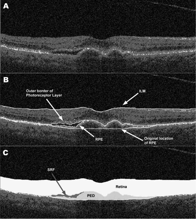
Methods: Best-corrected Snellen visual acuity was recorded for each patient. Raw exported StratusOCT images for each patient were analyzed using publicly available custom software entitled "OCTOR," which allows the precise positioning of prespecified boundaries on individual B-scans. Thickness and volume were calculated for morphologic parameters of interest: neurosensory retina, subretinal fluid, subretinal tissue (SRT), and pigment epithelial detachment.
Main outcome measures: OCT-derived measurements of retinal morphology and visual acuity.
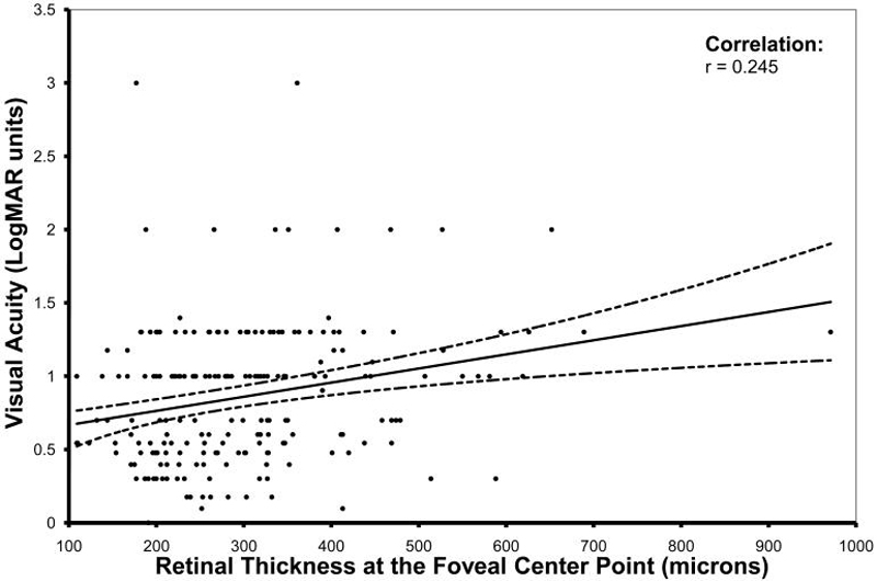
Results: An increased total volume of SRT was correlated with decreased visual acuity (r = 0.370, P<0.0001). Decreased visual acuity was also modestly correlated with increased thickness of the neurosensory retina at the foveal center point (r = 0.245, P = 0.0004). No statistically significant association was detected between visual acuity and the total volume of subretinal fluid or pigment epithelial detachment. The association between visual acuity and both the neurosensory retina and the SRT was stronger for lesions classified as minimally classic or occult on fluorescein angiography. For occult lesions, 20% of the variation in visual acuity could be predicted by a multiple regression model that incorporated age and SRT volume, whereas, for minimally classic lesions, 62% of the variation in visual acuity could be predicted by a multiple regression model that incorporated age, total neurosensory retinal volume, and total SRT volume.
Conclusions: The presence of increased SRT thickness and volume on OCT, and to a lesser extent increased neurosensory retinal thickness and volume, is associated with decreased visual acuity in neovascular AMD. However, because of the complex pathophysiology of neovascular AMD and, in part, the limitations of StratusOCT, these factors only account for a small degree of the variation in visual acuity that these patients exhibit. The detection of stronger correlations between retinal anatomy and visual acuity is likely to require the use of more advanced imaging modalities.
Financial disclosure(s): Proprietary or commercial disclosure may be found after the references.
Figures
References
-
- Rosenfeld PJ, Brown DM, Heier JS, et al. Ranibizumab for neovascular age-related macular degeneration. N Engl J Med. 2006;355:1419–31. - PubMed
-
- Avery RL, Pieramici DJ, Rabena MD, et al. Intravitreal bevacizumab (Avastin) for neovascular age-related macular degeneration. Ophthalmology. 2006;113:363–72. - PubMed
-
- Voo I, Mavrofrides EC, Puliafito CA. Clinical applications of optical coherence tomography for the diagnosis and management of macular diseases. Ophthalmol Clin North Am. 2004;17:21–31. - PubMed
-
- Kaiser PK, Blodi BA, Shapiro H, et al. Angiographic and Optical Coherence Tomographic Results of the MARINA Study of Ranibizumab in Neovascular Age-Related Macular Degeneration. Ophthalmology. 2007;114:1868–75. - PubMed
-
- Fung AE, Lalwani GA, Rosenfeld PJ, et al. An optical coherence tomography-guided, variable dosing regimen with intravitreal ranibizumab (Lucentis) for neovascular age-related macular degeneration. Am J Ophthalmol. 2007;143:566–83. - PubMed
Publication types
MeSH terms
Grants and funding
LinkOut - more resources
Full Text Sources
Medical
