Case Reports
. 2009 Feb;13(1):36-44.
doi: 10.1016/j.jaapos.2008.07.008. Epub 2008 Oct 18.

"Heavy Eye" syndrome in the absence of high myopia: A connective tissue degeneration in elderly strabismic patients

Affiliations
Case Reports

"Heavy Eye" syndrome in the absence of high myopia: A connective tissue degeneration in elderly strabismic patients

Tina Rutar et al. J AAPOS. 2009 Feb.

Abstract

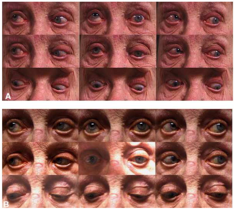
Purpose: In axial high myopes with "heavy eye" syndrome, orbital MRI can be use to demonstrate degeneration of the lateral rectus-superior rectus (LR-SR) band, with the result that the lateral rectus muscle slips inferiorly and causes esotropia and hypotropia. We investigated whether this degeneration might also cause strabismus in nonmyopic elderly patients.

Methods: Three elderly patients with strabismus, 3 strabismic high myopes, and 12 orthotropic elderly subjects underwent ophthalmic examinations and orbital MRI. The lateral rectus muscle position was determined relative to globe center from quasicoronal images and correlated with LR-SR band structure. MRI scans were compared with histology of 4 cadaveric orbits ranging in age from 17 months to 93 years.

Results: Two strabismic patients exhibited hypotropia; one exhibited esotropia. Mean axial length was 24.1 +/- 0.8 mm (mean +/- SD), compared with 31.6 +/- 1.4 mm for myopes. The lateral rectus muscle position of elderly strabismic subjects averaged 4.6 +/- 1.7 mm inferior to globe center, which was significantly lower than that of orthotropic elderly subjects (2.1 +/- 1.9 mm; p = 0.01) and similar to that of high myopes (5.1 +/- 3.2 mm). On MRI scanning, 100% of strabismic elderly orbits, 67% of strabismic myopic orbits, and 12.5% of control elderly orbits showed LR-SR band thinning, discontinuity, or displacement. LR-SR band degeneration was present histologically only in older cadavers.

Conclusions: Age-related LR-SR band degeneration permits the lateral rectus muscle to slip inferiorly in elderly nonmyopes, a mechanism of strabismus similar to myopic "heavy eye" syndrome. Imaging may assist in diagnosing this mechanical cause of age-related strabismus.


Figures

Fig. 1
Fig. 1. Diagnostic gaze positions of elderly strabismic subject 1, who underwent surgical repair of the inferiorly displaced left lateral rectus muscle
A - Preoperative photos show a –4 supraduction deficit of the left eye and a large left hypotropia in sursumversion. Note left upper eyelid ptosis and elevated eyelid crease. The right pupil was surgically mydriatic. B – Postoperative photos show improved supraduction with smaller left hypotropia in sursumversion.
Fig. 2
Fig. 2. Preoperative and postoperative Hess screen plots of elderly strabismic subject 1, showing left eye position with right eye fixating
A – Preoperative: There is an incomitant left hypotropia increasing in sursumversion. B – Postoperative: After surgical repair of the inferiorly displaced lateral rectus muscle, the left hypotropia improved.
Fig. 3
Fig. 3. Quasicoronal T1-weighted orbital MRIs of left orbits showing LR-SR bands and positions of the four rectus muscles relative to the globe
A - Normal elderly control. In this 74 year-old male, the lateral rectus muscle was 2.2 mm inferior to globe center and was oriented nearly vertically. The LR-SR band was thick, continuous and nondisplaced. Axial length was 24.2 mm. B - Elderly strabismic subject 1. In this 85 year-old female, the lateral rectus muscle was 7.6 mm inferior to globe center and was oriented obliquely rather than vertically. The LR-SR band was superotemporally displaced. Axial length was 23.3 mm. C - Strabismic myopic subject 1. In this 63 year-old female, the lateral rectus muscle was 7.4 mm inferior to globe center and was obliquely oriented. The LR-SR band was thin and superotemporally displaced, but immediately adjacent to the enlarged globe. Axial length was 30.5 mm. LPS/SR – levator palpebrae superioris/superior rectus, LR-SR – lateral rectus – superior rectus band, LR - lateral rectus, IR – inferior rectus, MR – medial rectus.
Fig. 4
Fig. 4
Intraoperative photo of left orbit of elderly strabismic subject 1 (left eye in adduction, surgeon’s view) showing absence of normal lateral rectus pulley connective tissues. The conjunctiva and Tenon’s are reflected temporally with a retractor and hook. Though no dissection of connective tissues was performed, there is prolapse of fat (arrow) into the surgical field. A small hook was used in an attempt to engage the connective tissues, which are absent. The thin line marks the lateral rectus muscle insertion.
Fig. 5
Fig. 5. Examples of LR-SR band degeneration in elderly strabismic subjects visualized by T2-weighted orbital MRI
A, B - Right and left orbits of elderly strabismic subject 3, showing thinned and superotemporally displaced LR-SR bands. C, D - Right and left orbits of elderly strabismic subject 2, showing severely thinned and discontinuous LR-SR bands. Only fragments of the connective tissue are visible.
Fig. 6
Fig. 6
Inferior displacement of lateral rectus muscle in strabismic elderly eyes compared to elderly control eyes and strabismic highly myopic eyes.
Fig. 7
Fig. 7. Histological sections of whole orbits stained with Masson’s trichrome stain, showing progressive thinning and superotemporal displacement of the LR-SR band (black arrow) with age. 10 micron thick sections
A - LR-SR band is thick and substantial in 17 month-old subject. B - LR-SR band is thick and substantial in 4 year-old subject, C - LR-SR band is thin and bulges superotemporally in 57 year-old subject. D - LR-SR band is very thin, is virtually indistinguishable from the lateral levator aponeurosis, and bulges superotemporally in 93 year-old subject, The lateral rectus muscle has also shifted inferiorly as observed in the current patients using MRI. LLA (green arrow) – lateral levator aponeurosis, LPS – levator palpebrae superioris, SR –superior rectus, LG – lacrimal gland, LR- lateral rectus, IO – inferior oblique.

References

    1. Demer JL. Pivotal role of orbital connective tissues in binocular alignment and strabismus: the Friedenwald lecture. Invest Ophthalmol Vis Sci. 2004;45:729–38. - PubMed
    1. Demer JL. Current concepts of mechanical and neural factors in ocular motility. Curr Opin Neurol. 2006;19:4–13. - PMC - PubMed
    1. Demer JL. Mechanics of the orbita. Dev Ophthalmol. 2007;40:132–57. - PMC - PubMed
    1. Hakim OM, El-Hag YG, Maher H. Persistence of eye movement following disinsertion of extraocular muscle. J AAPOS. 2008 in press. - PubMed
    1. Demer JL. More respect for connective tissues. J AAPOS. 2008 in press. - PubMed

Publication types